Treatment Overview

PDA closure surgery in India is a procedure used to close a patent ductus arteriosus (PDA)—a blood vessel that normally connects the aorta and pulmonary artery during fetal life but is expected to close after birth. When the duct remains open and causes a significant abnormal flow of blood, closing it can reduce the extra workload on the heart and lungs.
For many children and adults, PDA can now be closed through a minimally invasive transcatheter procedure, in which a small occlusion device is delivered through a blood vessel. Surgical ligation remains important when catheter closure is unsuitable, particularly in selected small or premature infants and in patients with unusual duct anatomy.
This guide explains PDA closure in India in detail—including symptoms, diagnosis, who needs closure, device closure versus surgery, preparation, procedure, recovery, risks, cost, hospital stay, follow-up and considerations for international patients.
Ventricular holes sit on Ventricular Septal Defect (VSD) Surgery in India. Aortic narrowing sits on Coarctation Repair Surgery in India. Broader surgical valve lists sit on Heart Valve Replacement Surgery in India. Pacing after heart block sits on Pacemaker Implantation in India. There is no live GAF pediatric-cardiac-surgery-only treatment page. This page is the named PDA-closure product. TOF lists sit on TOF Repair Surgery in India. Glenn lists sit on Glenn Procedure Surgery in India. Fontan lists sit on Fontan Procedure Surgery in India. Arterial-switch lists sit on Arterial Switch Operation in India.
GAF Healthcare planning for PDA closure is $3,500–$8,500 (typically 3–8 nights; parent stay expected). US comparison is $20,000–$60,000. Neighbouring congenital heart surgery is $8,000–$28,000 (typically 7–21 nights) when a broader congenital list is the honest product. Neighbouring VSD closure is $4,500–$11,000. Neighbouring ASD closure is $4,000–$9,500. Neighbouring coarctation repair is $6,000–$15,000. Neighbouring TOF repair is $6,500–$16,000. Neighbouring Glenn is $8,000–$18,000. Neighbouring Fontan is $9,000–$22,000. Neighbouring arterial switch is $12,000–$26,000. These are planning ranges from partner hospital cost sheets, not hospital quotations.
International patients comparing pediatric cardiac surgeons commonly start with Delhi NCR, Mumbai, Bengaluru, Chennai and Hyderabad. Partner pediatric cardiac surgery hospitals in Delhi NCR, Mumbai and Bengaluru, and in Chennai and Hyderabad, are a typical first filter because PDA closure needs echocardiography, catheter-lab or neonatal surgical backup and a pediatric or neonatal ICU when the infant is premature. City cost sheets include Delhi NCR, Mumbai, Bengaluru, Chennai and Hyderabad. Kolkata, Pune, Ahmedabad, Chandigarh and Kochi may have congenital theatres. They are not live GAF catalog cities on this site.
Important: A premature or collapsing infant with poor feeding, fast breathing or shock belongs in a local emergency department first. WhatsApp at +91 90443 46292 is for planned record review after the child is stable, not for neonatal collapse.
What is patent ductus arteriosus?
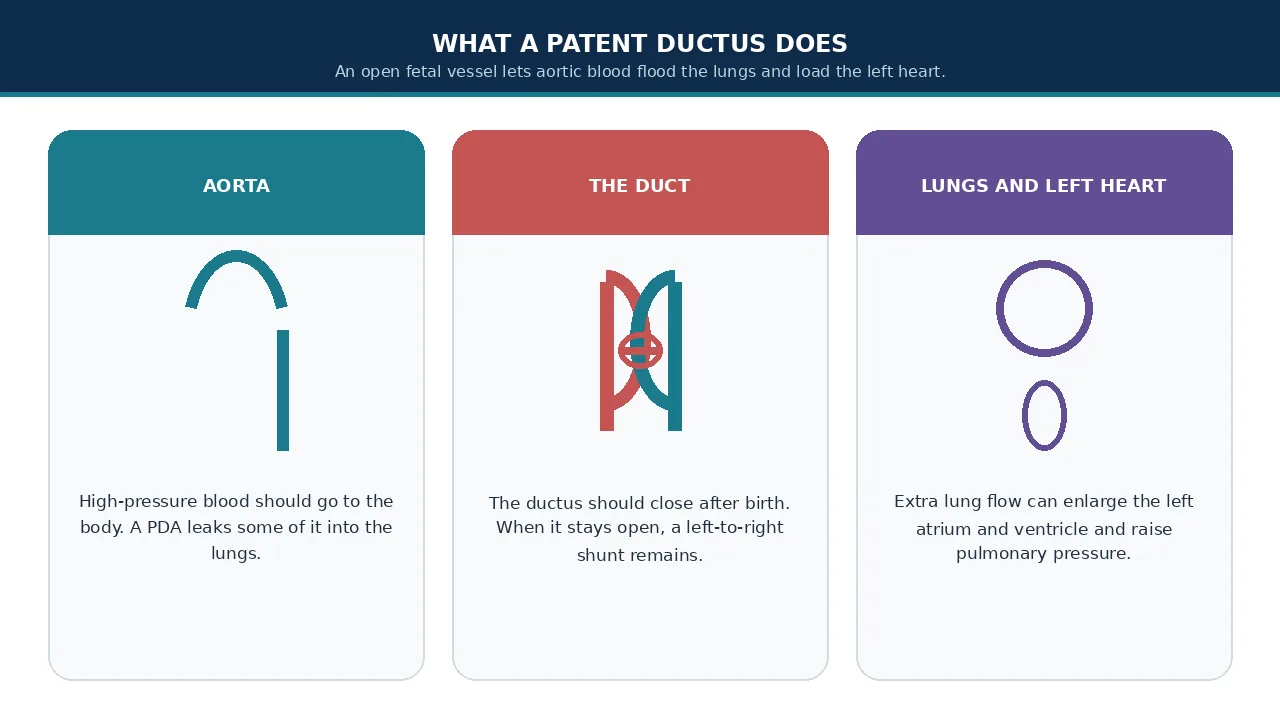
The ductus arteriosus is a normal fetal blood vessel connecting the pulmonary artery to the aorta.
Before birth, a baby does not use the lungs for oxygen exchange. The ductus therefore allows blood to bypass the fetal lungs.
After birth, the lungs begin functioning and the ductus normally constricts and eventually closes. Functional closure generally occurs early after birth, with anatomical closure occurring over the following weeks.
If the duct remains open, it is called patent ductus arteriosus, or PDA.
Because pressure in the aorta is normally higher than pressure in the pulmonary artery, blood can flow from the aorta into the pulmonary circulation. This is known as a left-to-right shunt.
A small PDA may cause little or no physiological effect. A larger PDA can send excessive blood to the lungs and increase the workload on the heart.
What happens if a PDA is left open?
The effect of PDA depends largely on its size and the patient's pulmonary vascular resistance.
A significant PDA can cause:
- Increased blood flow to the lungs
- Enlargement of the left atrium and left ventricle
- Breathing difficulties in infants
- Poor feeding or poor weight gain
- Heart failure in severe cases
- Pulmonary hypertension
- Progressive pulmonary vascular disease
- Rarely, irreversible changes in pulmonary circulation
The American Heart Association notes that untreated excessive pulmonary blood flow can eventually contribute to pulmonary hypertension and damage to the pulmonary blood vessels.
In adults, the decision to close a PDA becomes particularly dependent on the direction and magnitude of the shunt, pulmonary vascular resistance and evidence of cardiac volume overload. Current adult congenital-heart guidance specifically emphasizes assessing pulmonary hypertension before deciding on closure.
PDA closure surgery: when is it needed?
Not every PDA needs immediate closure.
The cardiologist evaluates:
- Size of the PDA
- Shape and anatomy of the duct
- Amount of blood shunting through it
- Heart chamber size
- Pulmonary artery pressure
- Symptoms
- Age and weight
- Prematurity
- Associated congenital heart defects
- Evidence of pulmonary vascular disease
A hemodynamically significant PDA is more likely to require intervention.
Indian consensus guidance has historically recommended individualized treatment according to PDA size, symptoms, pulmonary pressure and patient weight, with device closure favored in many patients above 6 kg when anatomy is suitable.
Important: A PDA associated with severe irreversible pulmonary vascular disease or a predominantly right-to-left shunt may not be suitable for closure. Closing a PDA in the wrong physiological setting can be harmful. This is why pulmonary pressure and pulmonary vascular resistance must be evaluated before intervention in selected patients.
Who may need PDA closure?
PDA closure may be considered in:
Babies and children
- Large or moderate PDA
- Symptoms of heart failure
- Poor growth
- Excessive pulmonary blood flow
- Enlargement of the left heart
- Recurrent respiratory problems associated with significant PDA
- A clinically significant PDA detected during echocardiography
Adults
An adult with PDA may require closure when there is a significant left-to-right shunt and evidence that the PDA is affecting cardiac structure or circulation.
The 2025 ACC/AHA adult congenital heart disease guidance recommends closure in appropriately selected adults with PDA-associated left-heart enlargement, net left-to-right shunting and no significant pulmonary vascular disease. It advises against closure when severe pulmonary hypertension and right-to-left shunting make closure unsafe.
Associated ventricular holes sit on VSD surgery. Associated aortic narrowing sits on coarctation repair. ASD lists sit on ASD Closure Surgery in India.
Ask whether device or ligation is the honest product
Types of PDA closure in India
There is no single closure technique that is appropriate for every patient.
The main treatment approaches are:
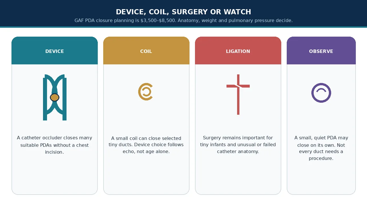
- Transcatheter PDA device closure
- Coil closure in selected small ducts
- Surgical PDA ligation or division
- Individualized care for premature infants, which may start with medicine rather than a procedure
The patient's age, weight, duct anatomy and pulmonary pressure are particularly important when choosing between these approaches.
Transcatheter PDA device closure
This is the most common minimally invasive approach when the patient's anatomy and size are suitable.
A catheter is introduced through a blood vessel, commonly in the groin. The cardiologist guides the catheter to the PDA and positions a specially designed occlusion device across the duct.
The device expands and blocks abnormal blood flow.
Over time, tissue grows around the device and provides permanent closure.
The American Heart Association describes catheter-based PDA closure as a common treatment option in children and adults.
Advantages
- No chest incision
- No surgical ligation in suitable patients
- Usually shorter recovery
- Small vascular access site
- Often short hospital stay
- Suitable for many children and adults
Surgical PDA ligation or division
Surgical closure may be recommended when catheter closure is not appropriate.
The surgeon accesses the PDA through the left side of the chest and closes it using a clip, stitches or division techniques depending on the anatomy and clinical situation.
Surgical closure may be particularly relevant in:
- Very small or premature infants
- PDA anatomy unsuitable for available devices
- Very large or unusual duct anatomy
- Associated conditions requiring surgery
- Failed or unsuitable catheter closure
- Selected ductal aneurysm or complicated PDA
Surgical PDA closure can be performed through conventional approaches or, in selected centers and patients, minimally invasive techniques such as thoracoscopic surgery.
Surgical PDA ligation is generally not an intracardiac open-heart operation. The surgeon closes the ductus rather than opening the heart chambers. It is still major pediatric cardiovascular surgery.
PDA closure in premature babies
PDA management in premature infants is different from management in older children.
Some premature babies may have spontaneous ductal closure or can initially be managed conservatively.
When a premature infant has a symptomatic or hemodynamically significant PDA, medication such as ibuprofen, indomethacin or acetaminophen/paracetamol may be considered depending on the infant's clinical circumstances.
If significant PDA persists despite medical treatment, or medication cannot be used, procedural closure may be considered.
Recent neonatal guidance emphasizes that procedural closure in extremely preterm infants should be individualized and considered particularly in selected infants with a confirmed large hemodynamically significant PDA and ongoing clinical problems.
PDA closure in a premature baby is not simply a smaller version of an adult procedure.
The baby's:
- Gestational age
- Birth weight
- Current weight
- Respiratory support
- Lung condition
- Kidney function
- Neurological condition
- PDA anatomy
- Pulmonary pressures
all influence treatment planning.
For international families, a premature or duct-dependent infant should generally be assessed and stabilized locally before anyone considers medical travel.
Ask about premature-infant suitability
PDA device closure procedure: step by step
The exact laboratory plan varies with the patient's anatomy.
A simplified catheter pathway is:
Step 1: Pre-procedure evaluation
The pediatric cardiologist reviews the child's medical history and performs a cardiovascular assessment.
Typical investigations may include:
- Echocardiography
- ECG
- Chest imaging when indicated
- Blood tests
- Oxygen saturation measurements
- Cardiac catheterization in selected cases
- Pulmonary pressure assessment when required
Echocardiography is particularly important for assessing PDA anatomy and its effect on the heart.
Step 2: Anesthesia
The type of anesthesia depends on the patient's age, size, clinical condition and institutional practice.
Young children commonly require general anesthesia or deep sedation to allow precise and safe device positioning.
Anesthesia planning becomes particularly important in premature or critically ill infants because respiratory and cardiovascular stability must be carefully maintained.
Step 3: Catheter insertion
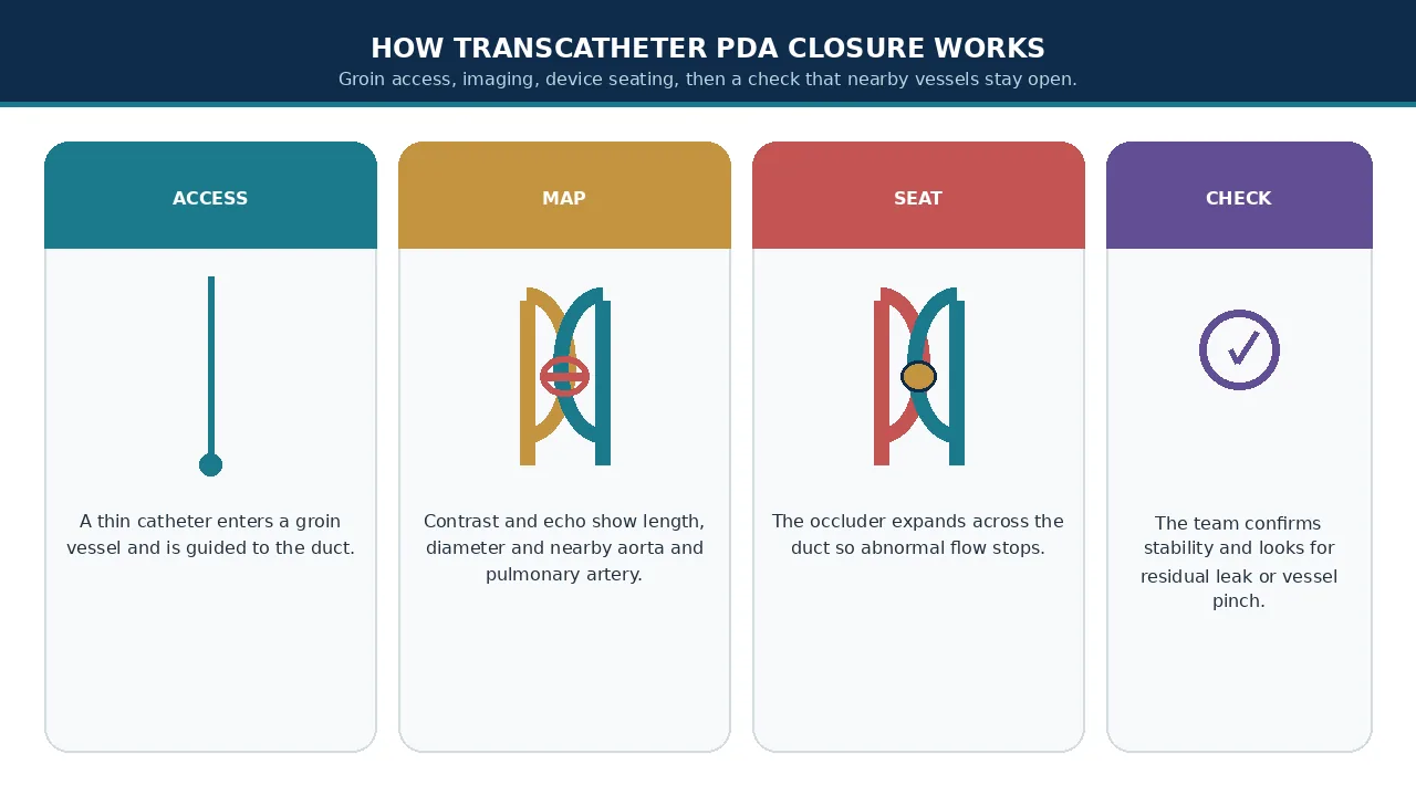
A thin catheter is introduced through a blood vessel, usually in the groin.
The catheter is carefully advanced toward the heart and great vessels.
Step 4: PDA imaging
Contrast imaging and echocardiographic guidance may be used to define:
- PDA length
- PDA diameter
- Shape
- Relationship with the aorta
- Relationship with the pulmonary artery
This information helps the cardiologist select the appropriate device.
Step 5: Device positioning
The PDA closure device is delivered through the catheter.
The cardiologist positions it so that it blocks the abnormal communication while minimizing obstruction of nearby vessels.
Step 6: Device release
Before releasing the device, the team checks its position and confirms that it is stable.
Imaging is used to evaluate blood flow and ensure that important adjacent structures are not obstructed.
Once the position is considered satisfactory, the device is released.
Step 7: Final assessment
The cardiologist checks for:
- Residual PDA flow
- Device stability
- Pulmonary artery obstruction
- Aortic obstruction
- Other procedural complications
The catheter is then removed.
What is a PDA closure device?
A PDA occlusion device is a small implant designed specifically to block blood flow through the persistent ductus.
Different devices are available for different PDA sizes, shapes and patient sizes.
One example used in appropriately selected patients is the Amplatzer Piccolo Occluder, which has been used for transcatheter PDA closure in small infants.
Device selection should never be based simply on the patient's age or weight. The anatomy of the PDA is a major consideration.
What is PDA coil closure?
Coil closure uses a small metallic coil to obstruct blood flow through the PDA.
It has historically been used particularly for smaller ducts.
With improvements in occluder technology, dedicated PDA devices are now used extensively in suitable patients.
The choice between a coil and an occluder device depends on duct anatomy, size, patient characteristics and the experience of the treating congenital-heart team.
PDA surgery vs device closure
| Feature | Device closure | Surgical ligation |
|---|---|---|
| Access | Blood vessel | Small chest incision |
| Chest incision | No | Yes |
| Heart opened? | No | No |
| General anesthesia | Often required in children | Usually required |
| Recovery | Usually quicker when uncomplicated | Usually longer |
| Suitable for premature infants | Selected cases | Important option in selected infants |
| Suitable for unusual PDA anatomy | Depends on anatomy/device | Can be useful |
| Hospital stay | Often short | Usually longer than uncomplicated catheter closure |
| Main risks | Device migration, vascular complications, residual shunt, obstruction | Bleeding, infection, nerve injury, respiratory complications and other surgical risks |
The table is a general clinical framework, not a treatment recommendation for an individual patient.
Transcatheter closure has increasingly become an important alternative to surgical ligation. Evidence suggests fewer cardiorespiratory complications and shorter hospitalization in appropriately selected patients, although patient selection strongly affects comparisons because the sickest and smallest infants may be more likely to require surgery.
How is PDA diagnosed?
Diagnosis begins with a clinical examination and echocardiography.
Echocardiography
An echocardiogram is usually the central investigation.
It can show:
- PDA location
- PDA size
- Direction of blood flow
- Pulmonary artery pressure estimates
- Left atrial size
- Left ventricular size
- Associated congenital heart defects
A hemodynamically significant PDA may produce left-sided chamber enlargement on echocardiography.
ECG
An ECG may provide supporting information about cardiac workload and rhythm.
Chest X-ray
A chest X-ray may be useful when assessing heart size or pulmonary blood flow, particularly in symptomatic patients.
Cardiac catheterization
Cardiac catheterization is not required for every PDA.
It may become important when:
- Pulmonary hypertension is suspected
- Pulmonary vascular resistance needs to be measured
- Anatomy is complex
- Closure suitability is uncertain
- An interventional procedure is planned
Adult congenital-heart guidelines recognize catheterization as useful when pulmonary hypertension is suspected in patients with PDA.
Symptoms of PDA
Symptoms vary considerably.
A small PDA may cause no noticeable symptoms.
A larger PDA can cause:
In infants
- Fast breathing
- Difficulty feeding
- Sweating while feeding
- Poor weight gain
- Frequent respiratory symptoms
- Tiredness
- Increased work of breathing
Severe infant distress belongs in a local emergency department, not on an international flight.
In older children
- Reduced exercise tolerance
- Breathlessness
- Easy fatigue
- Poor growth in significant cases
In adults
Some adults remain asymptomatic for years.
Others may develop:
- Breathlessness
- Reduced exercise tolerance
- Palpitations
- Fatigue
- Symptoms related to pulmonary hypertension or heart failure
Symptoms alone do not determine whether PDA closure is required. Echocardiography and physiological assessment are essential.
What are the risks of PDA closure?
PDA closure is generally well established, but no procedure is completely risk-free.
Risks of catheter/device closure
Potential complications include:
- Bleeding at the catheter site
- Groin hematoma
- Vascular injury
- Arrhythmia
- Residual PDA flow
- Device embolization or migration
- Obstruction of the descending aorta
- Left pulmonary artery obstruction
- Infection
- Thrombotic complications
- Rare cardiac or vascular injury
The likelihood varies considerably according to age, weight, PDA anatomy and the patient's underlying condition.
Risks of surgical PDA closure
Potential complications include:
- Bleeding
- Infection
- Respiratory complications
- Anesthesia-related complications
- Injury to nearby structures
- Chylothorax
- Recurrent laryngeal nerve injury
- Residual or recurrent flow
- Longer recovery compared with uncomplicated catheter closure
The risks can be substantially different in premature infants compared with healthy older children.
PDA closure success rate
PDA device closure has a high technical success rate when the patient's anatomy is suitable.
Published clinical literature reports approximately 96% procedural success for percutaneous closure in the broader population described in recent reviews, although success rates vary between patient groups and studies.
For families, the more useful question is not simply "What is the success rate?" but:
Is this particular PDA suitable for catheter closure, and what are the expected risks for this child?
A pediatric interventional cardiologist can answer that after reviewing the echocardiogram and other investigations.
Recovery after PDA device closure
After catheter closure, the patient is monitored for:
- Heart rate
- Blood pressure
- Oxygen saturation
- Catheter insertion site
- Heart rhythm
- Device position
- Residual blood flow
Many uncomplicated cases involve a relatively short hospital stay.
Children may experience mild discomfort at the groin access site for a short period.
Most children can gradually return to normal activity according to the treating cardiologist's instructions.
GAF planning is typically 3–8 nights, with parent stay expected. An uncomplicated older child may be toward the shorter end. Premature or complex infants can stay longer.
Recovery after surgical PDA ligation
Recovery following surgical closure is usually longer than uncomplicated catheter closure because the patient has a surgical incision.
The team monitors:
- Breathing
- Oxygenation
- Pain
- Wound healing
- Heart function
- Infection
- Fluid balance
Premature babies may need considerably longer intensive-care support because their overall medical condition—not only the PDA—can determine the length of hospitalization.
How long does PDA closure take?
The procedure time varies according to:
- Patient age
- Weight
- PDA anatomy
- Device selection
- Associated cardiac defects
- Whether additional procedures are required
A straightforward catheter closure may take roughly 1–3 hours, although the total time in the catheterization laboratory and hospital is longer because of anesthesia, preparation and recovery.
Surgical ligation may take longer and requires postoperative monitoring.
These are planning estimates, not fixed procedure times.
PDA closure cost in India
The cost of PDA closure in India varies significantly according to the procedure, hospital, city, device, patient age and weight, ICU requirements and whether the patient is an Indian or international patient.
GAF Healthcare planning for PDA closure is $3,500–$8,500 (typically 3–8 nights; parent stay expected). US comparison is $20,000–$60,000. Neighbouring broader congenital heart surgery is $8,000–$28,000. Published Indian rupee listings are not GAF quotations.
Indicative planning table
| Treatment pathway | Indicative GAF planning |
|---|---|
| PDA device or surgical closure | $3,500–$8,500; typically 3–8 nights |
| Neighbouring congenital heart surgery | $8,000–$28,000 when a broader congenital list is named |
| Neighbouring VSD closure | $4,500–$11,000 if a ventricular hole is added |
| Neighbouring ASD closure | $4,000–$9,500 on the named ASD sheet |
| Neighbouring coarctation repair | $6,000–$15,000 if arch narrowing is also named |
| Premature or ICU-heavy infant | May cost more because of intensive-care nights |
| International patient package | Written after echocardiography and weight review |
These are planning figures, not quotations.
The final estimate should be based on the patient's echocardiogram, current weight, pulmonary pressures, medical history and proposed treatment.
Request an itemised PDA estimate
What is included in the cost of PDA closure?
A hospital quotation may include some or all of the following:
- Interventional cardiologist or surgeon fees
- Anesthesia
- Catheter laboratory or operating-room charges
- The occlusion device or coil when used
- ICU charges
- Room charges
- Nursing
- Medicines
- Blood products when required
- Consumables
- Diagnostic tests
- Echocardiography
- Postoperative monitoring
- Follow-up consultation
However, inclusions vary by hospital.
International patients should ask for an itemized written quotation rather than relying only on a headline package price.
Ask whether the price includes the device, extra ICU nights, parent stay, residual-leak reintervention and complication-related expenses.
Additional costs international patients should consider
Families traveling to India may also need to budget for:
- Flights
- Visa-related expenses
- Accommodation
- Food
- Local transportation
- Companion expenses
- Additional investigations
- Longer ICU or hospital stay
- Additional procedures
- Medicines after discharge
- Follow-up imaging
- Emergency or unexpected treatment
For an infant or child, the cost of a parent or accompanying family member should be considered separately.
Why do families consider India for PDA closure?
India has established pediatric cardiology and congenital cardiac surgery programs.
Treatment may involve collaboration between:
- Pediatric cardiologists
- Pediatric interventional cardiologists
- Pediatric cardiac surgeons
- Adult congenital cardiologists
- Cardiac anesthetists
- Cardiac intensivists
- Neonatologists
- Radiologists
- Specialized nursing teams
The complexity of PDA makes the experience of the congenital heart team and availability of appropriate intensive care more important than simply choosing a hospital based on the advertised package price.
The AHA emphasizes that optimal congenital heart care depends on an integrated multidisciplinary team with lesion-specific expertise and perioperative care.
Choosing a hospital for PDA closure in India
When evaluating hospitals, families should ask:
1. Does the hospital treat newborn and premature congenital heart disease?
This is especially important for a small or premature infant.
2. Is a pediatric cardiac ICU or neonatal ICU available?
A dedicated unit can be important for postoperative or post-catheter monitoring.
3. Does the center provide both device closure and surgical ligation?
Having both options allows the team to change strategy if the anatomy is unsuitable for a device.
4. Is pediatric echocardiography and a catheter laboratory available?
Ask whether the center can perform:
- Pediatric echocardiography
- Cardiac catheterization
- Emergency surgical backup
- Pulmonary-pressure assessment when needed
5. Is follow-up echocardiography available?
PDA is not simply a one-time implant problem. Residual shunt and nearby-vessel flow need review.
6. Can the hospital manage associated congenital heart defects?
This is particularly important because PDA may occur together with VSD, coarctation or other abnormalities. Neighbouring VSD surgery and coarctation repair are different products.
Preparing for PDA closure in India as an international patient
International families should ideally send the patient's records before traveling.
Useful documents include:
- Echocardiogram reports
- Echo images or videos where available
- ECG
- Chest X-ray
- Blood test reports
- Previous hospitalization records
- Medication list
- Birth history
- Gestational age
- Current weight
- Oxygen requirement
- Previous cardiac procedures
- Previous surgical records
For premature infants, gestational age and current weight are particularly important.
The actual imaging is often more useful to the congenital team than a short written diagnosis.
Send echo, weight and pulmonary pressures before travel
Medical travel timeline for PDA closure
A typical international-patient pathway may look like this:
Step 1 — Send medical records
The family provides echocardiography and other available medical records.
Step 2 — Specialist review
A pediatric cardiologist, interventional cardiologist or congenital surgeon reviews the case.
Step 3 — Treatment plan
The team determines whether the patient may require observation, medical management, device closure, surgical ligation or additional evaluation.
Step 4 — Cost estimate
The hospital prepares a patient-specific quotation.
Step 5 — Travel planning
Once the patient is considered suitable for travel, the family can coordinate visa, flights and accommodation.
Step 6 — Hospital admission
Additional tests may be performed after arrival.
Step 7 — Treatment
Device closure or surgical ligation is performed according to the final clinical plan.
Step 8 — Recovery
The patient remains under hospital observation until medically stable.
Step 9 — Follow-up
The family receives a discharge plan and instructions for future echocardiography and cardiology review.
A coordinator can also open WhatsApp at +91 90443 46292 for planned records review.
How long should international patients stay in India?
There is no single stay duration for every PDA patient.
An older child undergoing uncomplicated catheter closure may need only a short period of hospital observation.
A premature infant may require prolonged hospitalization because of respiratory or other medical issues unrelated to the technical closure itself.
GAF planning is typically 3–8 nights, with parent stay expected. The travel plan should be based on the treating hospital's discharge assessment rather than a fixed number of days.
PDA closure follow-up
Follow-up generally includes clinical assessment and echocardiography.
The cardiologist may check:
- Device position
- Residual shunt
- Aortic flow
- Pulmonary artery flow
- Heart chamber size
- Pulmonary pressure
- Overall cardiac function
Indian consensus guidance has recommended clinical evaluation, ECG and echocardiography during the first year after uncomplicated PDA closure, with longer follow-up determined by residual defects or pulmonary hypertension.
Follow-up schedules can differ depending on the patient's age, procedure, associated heart disease and local protocol.
Ask about residual shunt and follow-up
Dental care after PDA closure
Families should maintain good oral and dental hygiene.
Historically, Indian congenital-heart guidance recommended infective endocarditis prophylaxis for six months after device or surgical closure in appropriate patients, while emphasizing ongoing dental hygiene.
However, antibiotic prophylaxis recommendations can vary according to the procedure, residual defect and current guideline used by the treating cardiologist.
Do not start antibiotics for dental procedures without specific advice from the patient's cardiologist or dentist.
PDA closure in adults
PDA is not exclusively a childhood condition.
Some adults are diagnosed incidentally after a murmur, echocardiogram or evaluation for breathlessness.
The adult evaluation is more complex when pulmonary hypertension is present.
Before closing an adult PDA, doctors may need to establish:
- Direction of the shunt
- Magnitude of the shunt
- Left-heart enlargement
- Pulmonary artery pressure
- Pulmonary vascular resistance
- Left ventricular function
The 2025 ACC/AHA adult congenital-heart guideline recommends evaluation for pulmonary arterial hypertension in adults with unrepaired PDA and uses pulmonary vascular resistance and shunt physiology to guide closure decisions.
Adults comparing adult congenital cardiology should still use the named pediatric or congenital PDA sheet when the product is PDA closure, not a generic adult CABG list. Neighbouring CABG is a different product.
Can every PDA be closed with a device?
No.
Device closure requires anatomy that can be safely treated with an available occluder.
Factors that may make device closure unsuitable include:
- Very unusual PDA shape
- Very large duct
- Inadequate device landing zone
- Extremely small patient size
- Proximity to important vessels
- Certain aneurysmal or complicated anatomy
- Severe pulmonary vascular disease
In these situations, surgical treatment or another management strategy may be considered.
Can a PDA close on its own?
Some small PDAs, particularly in infants, may close spontaneously.
The likelihood depends on age, prematurity and the characteristics of the duct.
Therefore, a newly diagnosed PDA does not automatically mean that the child needs an immediate procedure.
The cardiologist determines whether observation is reasonable or whether the PDA is producing enough physiological effect to warrant closure.
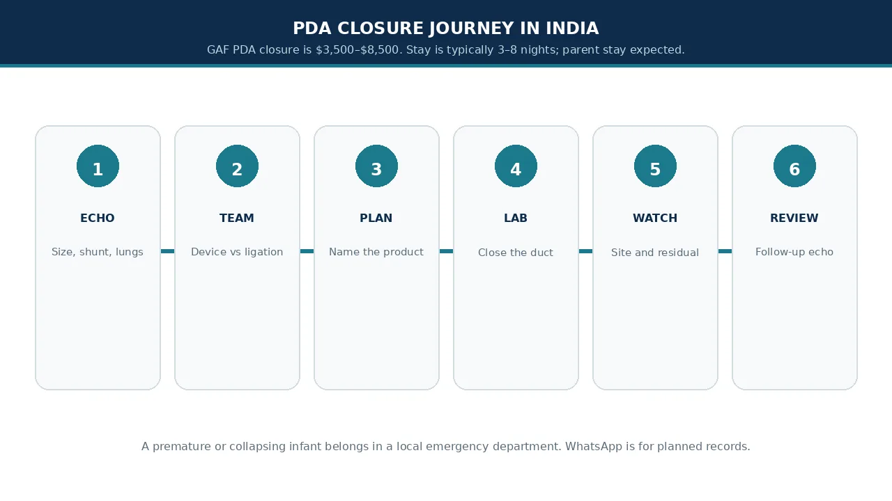
PDA closure in India: a typical patient journey
Step 1 — Medical records review. The family sends echocardiography and other available medical records.
Step 2 — Pediatric cardiologist assessment. The cardiology team determines whether the PDA is clinically significant.
Step 3 — Treatment planning. The team decides between observation, medical management, device closure or surgical closure depending on the patient's condition.
Step 4 — Cost estimate. The hospital provides an individualized estimate after reviewing the case.
Step 5 — Pre-procedure assessment. Required investigations and anesthesia assessment are completed.
Step 6 — PDA closure. The selected catheter-based or surgical procedure is performed.
Step 7 — Monitoring. The patient remains under cardiac or intensive-care observation as clinically required.
Step 8 — Discharge. The family receives medication, wound or access-site instructions and follow-up recommendations.
Step 9 — Follow-up echocardiography. The cardiologist checks the result and confirms satisfactory closure.
Questions to ask the pediatric cardiologist before PDA closure
Parents can take these questions to the consultation:
- How large is my child's PDA?
- Is the PDA hemodynamically significant?
- Is the heart enlarged because of the PDA?
- Is pulmonary pressure normal?
- Can the PDA close naturally?
- Does my child need closure now?
- Is catheter closure possible?
- Which device would be used?
- Why is this device appropriate?
- Would surgery be safer in this particular case?
- What are the major procedural risks?
- How long will my child stay in the hospital?
- Will ICU care be required?
- What is the expected recovery period?
- What follow-up tests are needed?
- Could there be residual PDA flow?
- Will my child need activity restrictions?
- What will happen if the PDA is not closed?
- What is included in the hospital quotation?
- Is the quoted device cost included?
Frequently asked questions about PDA closure surgery in India
Is PDA closure surgery a major operation?
It depends on the technique.
Transcatheter PDA device closure is minimally invasive and does not require an incision in the chest.
Surgical PDA ligation is more invasive because it requires access through the chest, although the heart itself is generally not opened.
Is PDA closure painful?
Catheter closure generally causes little postoperative discomfort beyond the vascular access site.
Surgical closure may cause more discomfort because of the chest incision.
Children receive appropriate anesthesia and pain management during and after the procedure.
Is PDA closure safe for babies?
PDA closure can be performed safely in appropriately selected infants, but the risk profile is different in premature and very low-weight babies.
Extremely premature infants may have respiratory, neurological, kidney and other medical problems that affect procedural risk.
Can a premature baby undergo PDA device closure?
Yes, selected premature infants can undergo transcatheter closure.
However, the decision requires specialized pediatric interventional cardiology expertise because very small babies have different anatomical and physiological considerations.
How long does a child stay in hospital after PDA device closure?
An uncomplicated older child may require only a short admission.
Premature babies and children with associated medical problems may need considerably longer hospitalization. GAF planning is typically 3–8 nights.
Can PDA come back after closure?
A successfully occluded PDA generally remains closed.
Rarely, residual flow may be detected after the procedure. Follow-up echocardiography allows the cardiologist to identify and monitor this.
Will my child have a normal life after PDA closure?
Many children with an isolated PDA that is successfully treated can lead normal, active lives.
The long-term outlook also depends on whether the child has pulmonary hypertension or another congenital heart condition.
The American Heart Association notes that most people do not have complications after PDA repair.
Can adults undergo PDA closure?
Yes.
Adults with PDA can undergo catheter-based closure when their anatomy and pulmonary vascular physiology make closure appropriate.
The evaluation is particularly important if pulmonary hypertension is present.
Is PDA closure an open-heart surgery?
No, not usually.
Catheter-based PDA device closure does not require opening the heart.
Surgical PDA ligation also does not generally involve opening the heart chambers; the surgeon accesses and closes the ductus through the chest.
What is the difference between PDA ligation and PDA device closure?
PDA ligation closes the duct surgically using a clip, stitches or division.
PDA device closure uses a catheter to place an occlusion device inside the PDA.
The appropriate approach depends on the patient's age, weight, anatomy and clinical condition.
How much does PDA closure cost in India?
GAF Healthcare planning for PDA closure is $3,500–$8,500, typically 3–8 nights, with parent stay expected. US comparison is $20,000–$60,000. A personalized hospital quotation is required for an accurate figure.
What happens if PDA is not treated?
A small PDA may remain clinically insignificant.
A significant untreated PDA can cause excessive pulmonary blood flow, left-heart enlargement, heart failure or pulmonary vascular disease.
The risk depends on the size of the PDA and the patient's physiology.
What documents should an international patient send before coming to India?
Send echocardiography reports and images, current weight, gestational age if relevant, pulmonary-pressure notes, previous surgical records, discharge summaries and a medication list.
Is a second opinion useful before PDA closure?
For a congenital cardiac procedure, a second specialist opinion can be useful, particularly when the choice between observation, device closure and surgical ligation is not straightforward.
When should I go to an emergency department?
Poor feeding, grey or blue skin, severe breathing difficulty or shock in a baby belongs in a local emergency department, not in a WhatsApp message.
PDA closure surgery in India: key takeaway
PDA closure surgery in India can stop abnormal flow through a persistent fetal vessel and reduce the extra load on the heart and lungs.
For many suitable children and adults, transcatheter device closure is a minimally invasive option. Surgical ligation remains important for selected premature infants and unusual anatomy.
The most important step is a detailed evaluation by a pediatric cardiologist, interventional cardiologist and/or congenital cardiac surgeon.
GAF Healthcare planning for the named PDA-closure sheet is $3,500–$8,500.
For international patients, treatment planning should be based on the patient's actual echocardiogram, weight and pulmonary pressures rather than diagnosis alone.
And even after a successful closure, follow-up echocardiography remains important, particularly for residual shunt, nearby-vessel flow and associated congenital heart disease.
Share reports for a preliminary PDA estimate
Related GAF Healthcare pages
- Ventricular Septal Defect (VSD) Surgery in India
- Coarctation Repair Surgery in India
- Heart Valve Replacement Surgery in India
- Pacemaker Implantation in India
- CABG Surgery in India
- PDA closure cost
- Congenital heart surgery cost
- VSD closure cost
- Pediatric cardiac surgeons in India
- Pediatric cardiac surgery hospitals in India
ASD lists sit on ASD Closure Surgery in India. Use this page plus the named modality sheets.
Top 10 sources
- American Heart Association — Patent Ductus Arteriosus (PDA).
- ACC/AHA Adult Congenital Heart Disease Guideline — PDA evaluation and closure.
- Indian Guidelines for Common Congenital Heart Diseases — PDA intervention.
- Indian Revised Consensus Statement on Congenital Heart Disease — follow-up after PDA closure.
- NCBI StatPearls — Patent Ductus Arteriosus.
- NCBI StatPearls — Anesthesia for Patients With PDA.
- Journal of Perinatology — Procedural Closure of PDA in Preterm Infants.
- GAF Healthcare PDA closure cost sheet — PDA Closure (Patent Ductus Arteriosus).
- GAF Healthcare congenital heart surgery cost sheet — Congenital Heart Surgery.
- GAF Healthcare VSD and coarctation sheets — VSD Closure and Coarctation Repair.
Medical disclaimer
This page is intended for general medical education and healthcare-planning purposes. It does not diagnose PDA or replace an examination by a qualified pediatric cardiologist, interventional cardiologist or congenital cardiac surgeon.
The decision to observe, to close with a device or coil, or to ligate surgically must be made individually after echocardiography and, when needed, pulmonary-pressure review.
Treatment costs are planning ranges from partner hospital cost sheets. A premature, collapsing or duct-dependent infant belongs in a local emergency department.
Treatment Process
- 1
Share echo
The family provides echocardiography, current weight and pulmonary-pressure notes before anyone books travel.
- 2
Congenital team review
A pediatric cardiologist and interventional team review whether the case is observation, device, coil or ligation.
- 3
Name the product
The team writes device closure, coil or surgical ligation after anatomy and pulmonary-pressure review.
- 4
Itemized estimate
GAF PDA closure planning is $3,500–$8,500. Neighbouring congenital heart surgery is $8,000–$28,000.
- 5
Travel only if stable
Stable planned cases travel after records review. A premature or collapsing infant belongs in a local emergency department.
- 6
Repeat essential tests
The receiving unit confirms echo, weight, saturation and fitness after arrival.
- 7
Close the named duct
Device, coil or ligation proceeds only after the product is named.
- 8
Watch residual flow
Access site, residual shunt and nearby-vessel flow are watched before discharge.
- 9
Follow-up echo
The family leaves with an echocardiography plan and who will watch residual shunt after returning home.