Treatment Overview
Ventricular Septal Defect (VSD) surgery in India is a procedure used to close an abnormal opening between the heart's two lower chambers, called the ventricles. VSD is a congenital heart defect, meaning it is usually present from birth. Small VSDs may close naturally and may only require regular monitoring, while larger or hemodynamically significant defects may require surgical or catheter-based closure.
India has established pediatric cardiac centers equipped to diagnose and treat children and adults with congenital heart defects. Treatment may include observation, medicines to control symptoms, open-heart VSD repair, or transcatheter device closure depending on the size, location, anatomy of the defect, symptoms, pulmonary pressure, age and overall health of the patient.
For international patients, VSD treatment in India can involve the complete pathway from diagnostic evaluation and echocardiography to pediatric cardiac surgery, intensive care, rehabilitation and follow-up.
Valve lists sit on Heart Valve Replacement Surgery in India when an outlet VSD has damaged the aortic valve. Pacing after complete heart block sits on Pacemaker Implantation in India. Aortic wall tears are a different product and sit on Aortic Dissection Repair Surgery in India. There is no live GAF ASD-closure, PDA-closure, Tetralogy-of-Fallot or pediatric-cardiac-surgery-only treatment page. This page is the named VSD-surgery product. LVAD lists sit on Left Ventricular Assist Device (LVAD) Procedure in India.
There is no live GAF VSD-only cost sheet. Neighbouring congenital heart surgery is $8,000–$28,000 (typically 7–21 nights; parent stay expected). US comparison is $80,000–$250,000. Neighbouring heart valve replacement is $7,000–$18,000 (typically 8–16 nights) when a valve is named in the same sitting. Named aortic valve replacement is $7,000–$18,500 (typically 8–16 nights) when SAVR is added for VSD-related aortic leak. Neighbouring pacemaker implantation is $3,500–$9,000 (typically 1–3 nights) if complete heart block requires pacing. Neighbouring minimally invasive cardiac surgery is $8,000–$20,000 when a mini approach can honestly complete isolated work. Transcatheter VSD device closure has no live GAF sheet and is quoted after echo and device selection. These are planning ranges from partner hospital cost sheets, not hospital quotations.
International patients comparing cardiac surgeons commonly start with Delhi NCR, Mumbai, Bengaluru, Chennai and Hyderabad. Partner cardiac surgery hospitals in Delhi NCR, Mumbai and Bengaluru, and in Chennai and Hyderabad, are a typical first filter because VSD closure needs a pediatric or congenital cardiac ICU, echo and emergency backup. City cost sheets include Delhi NCR, Mumbai, Bengaluru, Chennai and Hyderabad. Kolkata, Pune, Ahmedabad, Chandigarh and Kochi may have congenital theatres. They are not live GAF catalog cities on this site.
Important: Severe breathing difficulty, bluish discoloration, poor feeding, extreme lethargy or fainting belongs in a local emergency department first. WhatsApp at +91 90443 46292 is for planned record review, not an acute infant emergency.
What is a ventricular septal defect?
A ventricular septal defect, commonly called a VSD, is an opening in the wall separating the right and left ventricles of the heart.
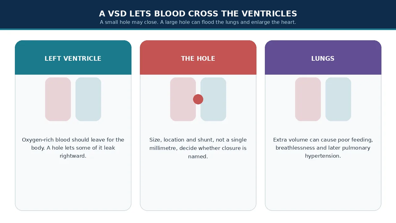
The left ventricle normally pumps oxygen-rich blood into the body's circulation, while the right ventricle pumps blood toward the lungs. When a VSD is present, blood can pass from the higher-pressure left ventricle into the right ventricle and then toward the lungs.
A small opening may cause little or no disturbance. A larger VSD can send excessive blood to the lungs, increasing the workload on both the heart and pulmonary circulation. Over time, an untreated significant VSD can contribute to heart failure, pulmonary hypertension and other complications.
VSDs are among the congenital heart defects that may be detected in infancy, childhood or adulthood. Some are discovered because a doctor hears a characteristic heart murmur, while others are identified during an echocardiogram performed for symptoms or another reason.
How does a VSD affect the heart?
The effect of a VSD depends mainly on its size, location and the amount of blood flowing through it.
With a significant left-to-right shunt:
- Blood moves from the left ventricle toward the right ventricle.
- Additional blood reaches the lungs.
- Pulmonary blood flow increases.
- The heart has to handle a greater volume of blood.
- The left side of the heart may become enlarged.
- Persistent high pulmonary blood flow can eventually contribute to pulmonary vascular disease.
A small VSD may produce little physiological effect. A large VSD can cause symptoms early in infancy, particularly as pulmonary resistance falls after birth and more blood begins flowing toward the lungs.
This is why doctors do not decide on surgery simply by measuring the hole. The physiological effect of the VSD is equally important.
Types of ventricular septal defects
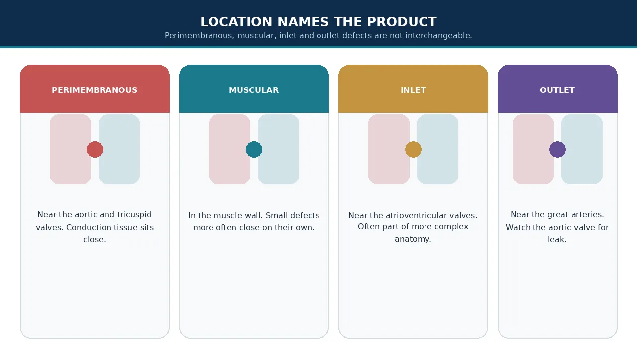
VSDs are classified according to their location in the ventricular septum.
Perimembranous VSD. A perimembranous VSD occurs near the membranous portion of the ventricular septum, close to the aortic and tricuspid valves. These defects require careful assessment because their location can be associated with nearby valve structures and the heart's electrical conduction system.
Muscular VSD. A muscular VSD is located within the muscular portion of the ventricular septum. Some muscular VSDs, particularly smaller defects, have a greater likelihood of spontaneous closure. Depending on anatomy, selected muscular VSDs may also be considered for catheter-based closure.
Inlet VSD. An inlet VSD is located closer to the area where blood enters the ventricles through the atrioventricular valves. Inlet defects can occur as part of more complex congenital heart conditions and therefore require detailed cardiac imaging.
Outlet or conoventricular VSD. These defects occur near the ventricular outflow tracts and the great arteries. Their relationship with the aortic and pulmonary valves is important because some defects can be associated with aortic valve problems.
The CDC describes these and other VSD types according to their anatomical location within the ventricular septum.
Ask which VSD type the echo is naming
What causes VSD?
In most cases, the exact cause of an isolated VSD is not known.
The defect develops while the baby's heart is forming during pregnancy. Genetic or chromosomal factors can contribute in some patients, and VSD may occur as part of a broader congenital heart syndrome.
VSD can also occur alongside other congenital heart conditions.
Importantly, parents should not assume that anything they did during pregnancy necessarily caused the defect. A congenital heart defect is a structural abnormality that develops during fetal heart formation, and the cause is often multifactorial or unknown.
Symptoms of ventricular septal defect
Symptoms vary considerably depending on the size of the VSD and the amount of blood passing through it.
Symptoms in babies. A baby with a significant VSD may develop rapid breathing, breathlessness, difficulty feeding, sweating during feeds, fatigue while feeding, poor weight gain, frequent respiratory symptoms, poor growth and signs of heart failure. A small VSD may cause no obvious symptoms and may only be detected because of a heart murmur.
Symptoms in older children. Older children may experience reduced exercise tolerance, breathlessness, easy fatigue, poor physical growth in some cases and recurrent respiratory symptoms.
Symptoms in adults. Some adults with a small VSD remain relatively asymptomatic. Others may experience shortness of breath during exertion, reduced exercise tolerance, fatigue, palpitations, a heart murmur, or symptoms related to pulmonary hypertension or valve disease.
Adults with congenital heart disease benefit from appropriate lifelong follow-up, particularly when the defect is moderate or complex or when there are residual problems after repair. The 2025 ACC/AHA/HRS/ISACHD/SCAI adult congenital heart disease guideline emphasizes specialized congenital-heart care and multidisciplinary assessment when appropriate.
Severe breathing difficulty, bluish discoloration, poor feeding, extreme lethargy or fainting is a local emergency department problem, not a WhatsApp message.
When does a VSD need surgery?
Not every VSD requires surgery.
Doctors generally consider closure when the VSD is causing a significant physiological problem or creates a meaningful risk of complications.
Possible reasons for closure include:
- A large VSD causing significant left-to-right shunting
- Heart failure symptoms despite medical management
- Poor growth or feeding difficulties
- Significant enlargement of the left ventricle
- Significant pulmonary overcirculation
- Pulmonary hypertension in an appropriate clinical setting
- Persistent hemodynamically significant shunting
- Certain VSD-related aortic valve problems
- Previous infective endocarditis associated with the VSD in selected patients
- Other anatomical complications
For adults, current congenital-heart guidelines use factors such as the size of the shunt, ventricular volume overload and pulmonary vascular resistance when deciding whether closure is appropriate. Closure is not appropriate in patients with severe irreversible pulmonary vascular disease in whom closing the VSD could be harmful.
The decision should therefore be individualized rather than based on the diameter of the VSD alone.
When may VSD surgery not be necessary?
A small VSD may not require an operation when the child has no significant symptoms, there is no important enlargement of the heart, pulmonary pressure is not concerning, the defect is expected to close spontaneously, there is no significant valve complication, and the cardiologist considers observation safe.
Many small VSDs close spontaneously, particularly during childhood. Regular echocardiographic follow-up allows the cardiologist to monitor the defect and determine whether intervention becomes necessary.
Diagnosis of ventricular septal defect
A VSD may be suspected during a physical examination or detected during prenatal imaging.
Echocardiography is the main diagnostic test for VSD. An echocardiogram can provide information about VSD location, size, direction of blood flow, shunt severity, heart chamber dimensions, ventricular function, pulmonary pressure estimates, heart valve function and associated congenital heart defects. It is usually the most important investigation for treatment planning.
Electrocardiogram. An ECG evaluates the heart's electrical activity and may provide evidence of chamber enlargement or rhythm abnormalities.
Chest X-ray. A chest X-ray may show heart enlargement or increased pulmonary vascular markings in patients with significant pulmonary blood flow.
Cardiac catheterization. Cardiac catheterization may be required in selected patients when doctors need more detailed information about pulmonary artery pressure, pulmonary vascular resistance, intracardiac pressures, oxygen saturation or complex anatomy. It can also be used for catheter-based VSD closure in selected cases.
Cardiac CT or MRI. Advanced imaging may be used when echocardiography does not provide enough anatomical information or when associated congenital abnormalities need further evaluation.
Send echocardiography images, not only the written report
Preparing for VSD surgery in India
Before surgery, the pediatric cardiology and cardiac surgery team typically reviews echocardiography, ECG, chest imaging when required, blood tests, oxygen saturation, weight and nutritional status, pulmonary pressure, ventricular function, associated heart defects, previous cardiac procedures, current medications, history of infections and any genetic or syndromic diagnosis.
For an international patient, the Indian hospital may review the medical records before travel.
Families should ideally send a recent echocardiogram report, echocardiogram images or video if available, ECG, CT/MRI reports if performed, previous discharge summaries, medication list, blood investigations, vaccination information for children where relevant and previous surgical records.
This allows the treating team to assess the case before the family travels to India.
VSD surgery options in India
The treatment approach depends on the patient's anatomy.
The main options are:
- Observation
- Medical management
- Open surgical VSD closure
- Transcatheter VSD closure
- Staged treatment in selected complex cases
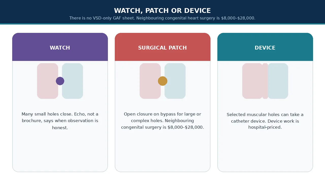
Open VSD closure surgery
Open-heart surgery is the standard approach for many significant VSDs.
During surgery, the patient receives general anesthesia and is connected to a heart-lung bypass machine.
The surgeon reaches the heart and closes the VSD, usually with a patch or, in selected situations, direct suturing.
The patch may be made from specialized surgical material or biological tissue. Over time, the body's own tissue grows over the patch.
Open repair remains particularly important for large VSDs, complex VSD anatomy, multiple VSDs, VSDs unsuitable for device closure, VSDs associated with other defects requiring surgery, and some defects close to important cardiac structures.
The American Heart Association notes that large VSDs may require surgical patch closure and that some VSDs can instead be closed in the cardiac catheterization laboratory.
Neighbouring congenital heart surgery is $8,000–$28,000.
Ask whether a patch or a device is the honest product
Transcatheter VSD closure
Selected VSDs can be closed without conventional open-heart surgery.
A catheter is introduced through a blood vessel, usually from the groin, and guided toward the heart. A closure device is positioned across the defect.
Once correctly positioned, the device blocks abnormal blood flow through the VSD.
This approach can be attractive in carefully selected patients because it avoids a traditional chest incision and cardiopulmonary bypass.
However, not every VSD is suitable for device closure.
The decision depends on VSD location, size, relationship to heart valves, distance from conduction tissue, patient age and size, associated heart defects, device availability and experience of the treating center.
Open surgery remains the preferred approach for many VSDs. There is no live GAF VSD-device sheet. A device list is quoted after echo and anatomy review.
VSD surgery vs device closure
| Feature | Surgical VSD closure | Transcatheter VSD closure |
|---|---|---|
| Main approach | Open-heart surgery | Catheter-based |
| Chest incision | Usually required | Usually avoided |
| Heart-lung machine | Usually required | Usually not required |
| Suitable for large/complex VSDs | Often | Limited |
| Suitable for selected muscular VSDs | Yes | Sometimes |
| Device required | No | Yes |
| Hospital recovery | Depends on age and complexity | Often shorter when uncomplicated |
| Neighbouring GAF sheet | Congenital heart surgery $8,000–$28,000 | Hospital-priced; no GAF device sheet |
The choice should be made by a congenital or pediatric cardiac team after reviewing the complete cardiac anatomy rather than simply choosing the least invasive option.
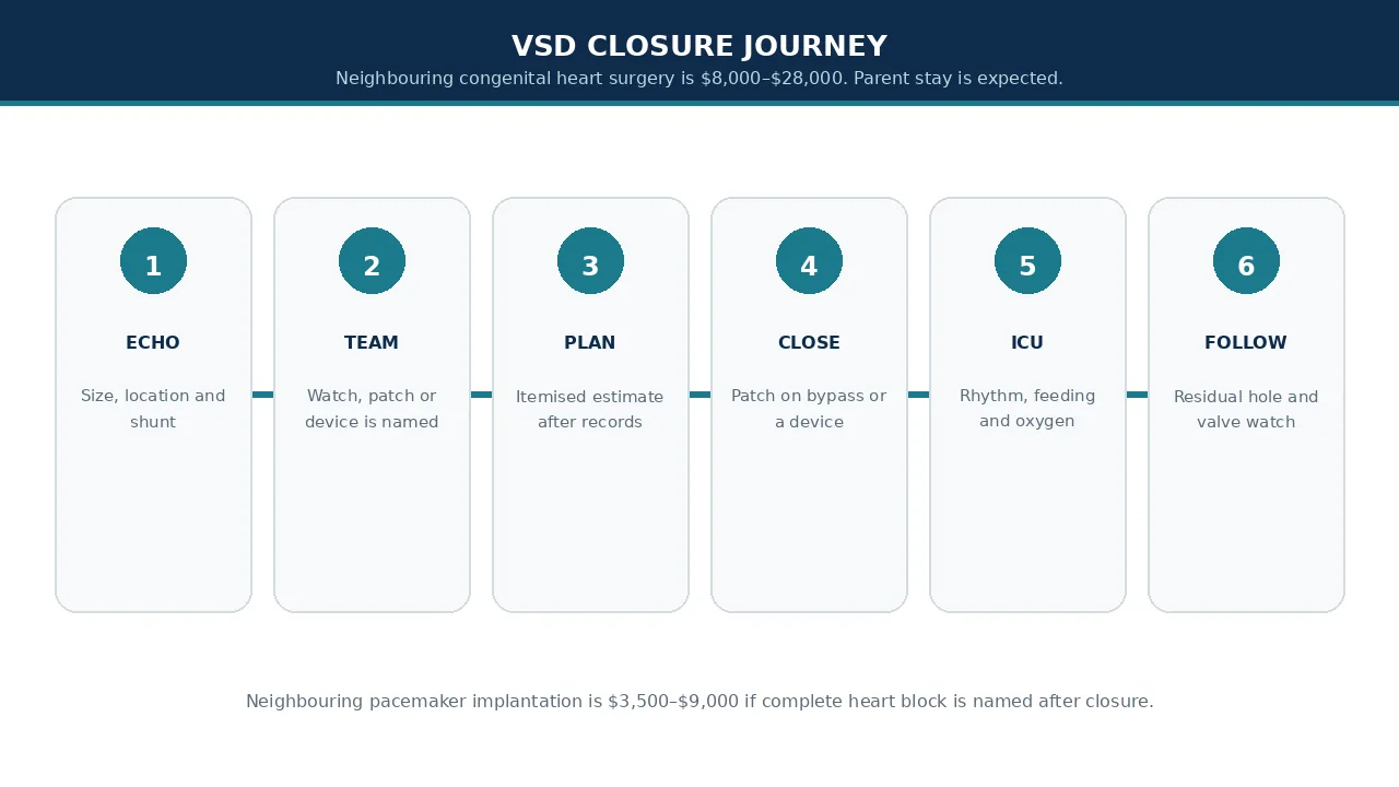
VSD surgery procedure: step by step
For an open surgical VSD repair, the process generally includes the following stages.
Step 1: General anesthesia. The patient is placed under general anesthesia.
Step 2: Surgical access. The cardiac surgeon obtains access to the heart through an appropriate surgical approach.
Step 3: Cardiopulmonary bypass. The patient is connected to a heart-lung machine, which temporarily performs the circulation and oxygenation functions of the heart and lungs.
Step 4: Identification of the VSD. The surgeon identifies the defect and evaluates nearby structures, including the heart valves and conduction system.
Step 5: Closure of the VSD. The defect is closed with a patch or sutures depending on its location and characteristics.
Step 6: Assessment of the repair. The surgical team checks the heart for residual shunting, valve function and other issues before completing the procedure.
Step 7: ICU recovery. The patient is transferred to the cardiac intensive care unit for close monitoring.
The precise operative technique varies according to the VSD's anatomy and whether other congenital heart problems require simultaneous correction.
How long does VSD surgery take?
The duration of VSD surgery varies.
A straightforward isolated VSD repair is different from a procedure involving multiple VSDs, complex congenital anatomy, valve repair, additional cardiac defects, very young infants or previous cardiac surgery.
Families should therefore avoid relying on a single standard operation time when planning travel or accommodation.
The hospital will generally provide an expected surgical and hospitalization timeline after reviewing the patient's medical records.
VSD surgery recovery
After surgery, the patient is usually monitored in a cardiac intensive care unit before moving to a regular ward.
Recovery can involve monitoring of heart rhythm, oxygen levels, blood pressure, breathing, fluid balance, heart function, surgical wound, feeding and nutrition.
Children may initially have reduced appetite or energy. Infants may require careful feeding and nutritional support.
Recovery time varies substantially with age and complexity. Neighbouring congenital heart surgery planning is typically 7–21 nights, with parent stay expected.
A child undergoing repair of an isolated VSD may recover considerably faster than a newborn undergoing repair of a large VSD combined with other congenital abnormalities.
Possible risks and complications of VSD surgery
VSD closure is a major cardiac procedure, and although modern congenital cardiac surgery has good outcomes, complications can occur.
Potential complications include bleeding, infection, abnormal heart rhythm, residual VSD or residual shunt, complete heart block, fluid accumulation, lung complications, heart failure, kidney complications, neurological complications, problems involving heart valves and need for further intervention.
Complete heart block and residual shunting are recognized complications of VSD repair.
The individual risk depends on age, weight, VSD location, VSD size, pulmonary pressure, heart function, associated congenital heart defects, previous surgery and overall health.
A surgeon should provide patient-specific risk estimates after reviewing the complete clinical picture.
Neighbouring pacemaker implantation is $3,500–$9,000 if complete heart block requires pacing.
Is VSD surgery safe?
VSD surgery has become an established treatment for significant defects, particularly when performed by experienced congenital cardiac teams.
However, “safe” does not mean risk-free.
For children with isolated VSD, published clinical references describe generally favorable outcomes, but individual risk varies significantly with anatomy and clinical condition.
Families should ask the treating center for outcomes relevant to the specific type and age group of the patient, rather than relying on a generic success percentage.
VSD surgery success rate
There is no single success rate that applies to every VSD patient.
Outcomes depend on type of VSD, size of the defect, patient age, weight, presence of pulmonary hypertension, associated congenital heart disease, ventricular function, timing of surgery and experience of the congenital cardiac center.
For a straightforward isolated VSD, complete closure and good long-term function are common outcomes.
The long-term outlook is generally favorable for children whose VSD is successfully closed and who do not have significant residual abnormalities.
VSD surgery cost in India
The cost of VSD surgery in India depends on the patient's age, complexity of the defect, surgical approach, hospital, ICU requirements, room category, investigations and whether additional cardiac procedures are required.
There is no live GAF VSD-only sheet. Neighbouring congenital heart surgery planning is $8,000–$28,000, typically 7–21 nights. Comparable US planning is $80,000–$250,000. Brochure rupee figures are not international self-pay quotations and are not used on this page.
| Neighbouring GAF sheet | Planning range | Typical stay |
|---|---|---|
| Congenital heart surgery | $8,000–$28,000 | 7–21 nights |
| Heart valve replacement | $7,000–$18,000 | 8–16 nights |
| Aortic valve replacement | $7,000–$18,500 | 8–16 nights |
| Minimally invasive cardiac surgery | $8,000–$20,000 | 5–12 nights |
| Pacemaker implantation | $3,500–$9,000 | 1–3 nights |
| Transcatheter VSD device | Hospital-priced; no GAF sheet | Quoted after echo |
A written package quotation should clearly state what is included and excluded: surgeon fees, cardiologist fees, anesthesia, operation theatre, heart-lung machine, ICU, room, medicines, investigations, blood products if required, medical devices, follow-up during hospitalization and treatment of complications.
Request an itemised VSD estimate
Factors that affect VSD surgery cost in India
Hospital. Different hospitals have different pricing structures, infrastructure and package models.
City. Live GAF catalog cities are Delhi NCR, Mumbai, Bengaluru, Chennai and Hyderabad.
Patient age. Neonatal and infant cardiac surgery can require highly specialized intensive care and monitoring.
Complexity of VSD. An isolated VSD is different from a VSD associated with other congenital heart defects.
Surgical vs device closure. Device-based closure involves the cost of the closure device and catheter laboratory procedure.
ICU stay. A prolonged ICU stay can increase the overall cost.
Complications. Additional treatment may increase the final bill.
Hospitals for VSD surgery in India
Families considering VSD surgery in India should focus on congenital and pediatric cardiac expertise, rather than choosing a hospital solely because it is a large general hospital.
Important factors include a dedicated pediatric cardiac surgery program, pediatric cardiology, congenital cardiac surgeons, pediatric cardiac anesthesia, pediatric cardiac ICU, neonatal intensive care capability, 24/7 emergency support, a cardiac catheterization laboratory, echocardiography expertise, experience with complex congenital heart disease, multidisciplinary care and long-term congenital heart follow-up.
Start with live GAF cardiac surgery hospitals in Delhi NCR, Mumbai, Bengaluru, Chennai and Hyderabad. Kolkata, Pune, Ahmedabad, Chandigarh and Kochi may have congenital theatres. They are not live GAF catalog cities on this site.
For a complex congenital heart case, the experience of the specific cardiac team and center may be more relevant than the hospital's general reputation.
Why international patients consider India for VSD treatment
India has developed specialized pediatric and congenital cardiac programs capable of treating patients ranging from infants to adults with congenital heart disease.
International patients may seek treatment in India because of specialized cardiac teams, advanced diagnostic facilities, pediatric cardiac ICUs, experience with congenital heart surgery, availability of surgical and catheter-based treatment, multidisciplinary care and international patient departments.
Treatment decisions should be based primarily on the patient's medical needs, the expertise of the treating center and the proposed treatment plan.
VSD surgery for international patients
International families should ideally complete a medical review before traveling.
- Send medical records. Echocardiogram, ECG, medical reports, previous surgery records, current medicines, blood tests, growth information and any CT/MRI reports.
- Receive medical opinion. The hospital's pediatric cardiologist or cardiac surgeon reviews the case.
- Obtain a treatment plan. Observation, medication, open surgery, device closure or additional investigations.
- Receive cost estimate. An itemized quotation after the product is named.
- Arrange travel. Travel should be planned after the medical team confirms that the patient is clinically fit to travel.
- Preoperative assessment in India. Repeat investigations after arrival.
- Treatment. The procedure is performed after the final medical assessment.
- Recovery and follow-up. The patient remains under medical supervision until the treating team considers discharge appropriate.
Share records before anyone books travel
WhatsApp coordination for planned travel is available at +91 90443 46292.
How long should international patients stay in India?
The total stay depends on the procedure, age, recovery and medical condition.
A medical traveler should consider time for initial consultation, diagnostic testing, preoperative assessment, surgery, ICU recovery, ward recovery, follow-up echocardiography, wound assessment and final clearance for travel.
Neighbouring congenital-heart planning is typically 7–21 nights, with parent stay expected. Families should avoid booking a return flight immediately after the expected discharge date.
The treating surgeon should provide travel advice before the patient leaves India.
VSD surgery in babies
VSD can have a particularly important impact on infants when the defect is large.
A significant VSD may cause rapid breathing, feeding difficulty, sweating during feeding, poor weight gain, heart failure and increased pulmonary blood flow.
Some infants can initially be managed with medicines and nutritional support while the medical team determines the appropriate timing of repair.
Large symptomatic VSDs may require repair during infancy to prevent ongoing complications.
The exact timing is individualized and depends on the child's anatomy, symptoms, pulmonary pressure, growth and response to medical treatment.
VSD surgery in older children
Older children may have VSDs that were previously undiagnosed, small and monitored, partially closed, or associated with another congenital heart problem.
The decision to close a VSD in an older child depends on its current physiological effect and potential long-term complications.
A child who is active and asymptomatic may not necessarily need surgery.
VSD surgery in adults
Some adults reach adulthood with an unrepaired VSD.
A small VSD may remain clinically stable, while a larger defect can cause left ventricular volume overload, pulmonary hypertension, aortic valve complications, arrhythmias, heart failure or other congenital-heart complications.
Adult VSD closure requires careful assessment of pulmonary vascular resistance and the direction and magnitude of the shunt.
Current adult congenital heart disease guidance recommends specialized assessment for adults with congenital heart disease, particularly when the anatomy or physiology is moderate or complex.
Can VSD close on its own?
Yes. Some small VSDs close spontaneously without surgery.
The likelihood depends on VSD size, location, age and anatomy. Muscular VSDs, in particular, may close spontaneously during childhood.
However, families should not assume that a VSD will close simply because it is small. Regular follow-up allows the cardiologist to determine whether the defect is becoming smaller and whether it is affecting the heart.
Medicines for VSD
Medicines do not close the hole.
They may be used to manage symptoms caused by significant VSDs, especially in infants who have heart failure or excessive fluid accumulation.
Depending on the clinical situation, treatment may include diuretics, other heart-failure medicines and nutritional support.
These treatments may stabilize the child while the cardiac team determines whether and when definitive closure is required.
Follow-up after VSD repair
VSD closure is not necessarily the end of cardiac care.
Follow-up may include clinical examination, echocardiography, ECG, assessment of heart rhythm, evaluation of ventricular function, monitoring of heart valves and assessment for residual shunting.
The American Heart Association recommends continued cardiology follow-up after VSD repair, with frequency determined by the patient's anatomy and clinical status.
Can a child live a normal life after VSD surgery?
Many children whose VSD has been successfully repaired go on to lead healthy, active lives.
Children with a small VSD or successfully repaired VSD often do not require permanent activity restrictions, although recommendations depend on the presence of residual defects, pulmonary hypertension, ventricular dysfunction, arrhythmias or other congenital heart conditions.
The cardiologist should determine when a child can return to school, sports, swimming, physical education, normal play and travel.
Exercise after VSD repair
Exercise recommendations depend on the patient's postoperative condition.
A child with complete VSD closure, normal heart function, no significant residual defect, no pulmonary hypertension and no significant arrhythmia may often be able to participate in normal physical activities.
Patients with residual cardiac problems may require individualized restrictions.
Do not use a generic internet exercise recommendation in place of the cardiologist's assessment.
Pregnancy after VSD repair
Women who had a VSD repaired successfully and have no significant pulmonary hypertension or other cardiac complications may often have a favorable pregnancy outlook.
However, pregnancy increases the workload on the cardiovascular system.
Women with repaired or unrepaired congenital heart disease should receive appropriate pre-pregnancy counseling, particularly if there is pulmonary hypertension, ventricular dysfunction, significant valve disease, residual shunting or complex congenital heart disease.
VSD and pulmonary hypertension
One of the important reasons doctors monitor a significant VSD is its effect on the pulmonary circulation.
A large untreated VSD can expose the pulmonary arteries to increased blood flow and pressure. Over time, severe pulmonary vascular disease can develop.
In advanced cases, pulmonary vascular resistance may become so high that the original left-to-right shunt changes direction, resulting in Eisenmenger syndrome.
Once severe irreversible pulmonary vascular disease has developed, VSD closure may no longer be appropriate and can be harmful.
This is one reason timely evaluation and appropriate follow-up matter.
VSD and aortic valve problems
Certain VSDs, particularly those located near the aortic valve, can affect the valve over time. Aortic regurgitation may develop in selected patients.
If a VSD contributes to progressive aortic valve leakage, surgical closure may be considered even when the VSD itself is not very large.
This is one reason that the location of a VSD matters as much as its size. Surgical valve lists sit on Heart Valve Replacement Surgery in India. Neighbouring aortic valve replacement is $7,000–$18,500.
VSD and infective endocarditis
Infective endocarditis is an infection involving the inner lining of the heart or heart valves.
The risk and need for preventive antibiotics depend on the patient's specific cardiac condition and whether the VSD has been repaired.
Current patient guidance from the American Heart Association indicates that most people with an unrepaired VSD do not routinely require antibiotic prophylaxis before dental procedures. After successful repair, prophylaxis may be recommended during the first six months of healing in appropriate situations, or longer when a residual defect remains adjacent to a repair.
Good dental hygiene and regular dental care remain important. Patients should follow the specific recommendations of their cardiologist.
Unexplained persistent fever after a repair belongs in a local emergency department.
Questions to ask the VSD surgeon
Before proceeding with treatment, families should consider asking:
- What type of VSD does my child have?
- How large is the VSD?
- Is the VSD causing significant left-to-right shunting?
- Is the heart enlarged?
- Is pulmonary pressure elevated?
- Could the VSD close naturally?
- Does my child actually need surgery now?
- Is catheter closure possible?
- Why do you recommend surgery instead of device closure?
- Which surgical technique will you use?
- Will any other heart defect be repaired during the same operation?
- What are the major risks in this particular case?
- How long will ICU care be required?
- How long is the expected hospital stay?
- What is the estimated total cost?
- What does the package include?
- Will blood products be required?
- What follow-up is needed after discharge?
- When can my child return to school and normal activities?
- When is it safe to travel home?
Send those answers with the echo
Documents to bring for VSD treatment in India
International patients should ideally carry both digital and printed copies of passport, previous medical reports, echocardiography reports, echocardiography images or videos, ECG, chest X-ray, CT/MRI reports, blood test reports, previous discharge summaries, operative reports, medication list, allergy information, vaccination records where relevant and previous genetic testing if available.
Choosing a VSD surgery center in India
The following checklist can help families compare centers objectively:
- Dedicated pediatric cardiologists experienced in congenital heart disease
- Congenital cardiac surgeons who routinely perform VSD repairs in the relevant age group
- Pediatric cardiac ICU
- Neonatal intensive care for newborns and very small infants
- Pediatric cardiac anesthesiologists
- Cardiac catheterization laboratory for selected device closure
- Multidisciplinary coordination
- Long-term congenital-heart follow-up after returning home
These factors are often more useful than simply comparing hospital brand names.
Frequently asked questions
Is VSD surgery necessary for every child? No. Small VSDs may close spontaneously or remain clinically insignificant and may only require periodic follow-up. Surgery is considered when the defect causes significant physiological effects or complications.
What is the most common treatment for a large VSD? For many large or significant VSDs, surgical patch closure is the established treatment. Selected defects can be closed using a catheter-based device.
Can VSD be treated without open-heart surgery? Yes, selected VSDs can be closed using a catheter and device. However, suitability depends heavily on the location and anatomy of the defect. There is no live GAF VSD-device sheet.
Can a VSD close naturally? Yes. Many small VSDs, particularly some muscular defects, may close spontaneously during childhood.
What happens if a large VSD is left untreated? A significant untreated VSD can result in excessive pulmonary blood flow, heart failure, pulmonary hypertension and progressive pulmonary vascular disease.
Is VSD surgery performed on babies? Yes. Infants with significant symptomatic VSDs may require repair during infancy. The timing depends on the baby's symptoms, growth, anatomy and pulmonary circulation.
What is the VSD surgery cost in India? There is no live GAF VSD-only sheet. Neighbouring congenital heart surgery is $8,000–$28,000 (typically 7–21 nights). Device closure is hospital-priced.
How long does a child stay in hospital after VSD surgery? Neighbouring congenital-heart planning is typically 7–21 nights. Infants and children with associated congenital heart conditions may require longer hospitalization.
Is VSD surgery painful? The operation itself is performed under general anesthesia. After surgery, children may experience discomfort, which is managed with appropriate pain-control medicines.
Can a child play normally after VSD surgery? Many children with successful repair and normal heart function can eventually participate in normal activities. Activity restrictions, if any, should be determined by the cardiologist.
Will my child need another operation? Most children with a successful isolated VSD repair do not require another operation, although a small number may require additional treatment for residual defects, valve problems or other complications.
Can adults undergo VSD closure? Yes. Adults with an unrepaired VSD may be considered for closure when the physiological and anatomical criteria are appropriate. Severe irreversible pulmonary vascular disease can make closure inappropriate.
Can VSD cause pulmonary hypertension? Yes. A large untreated VSD can increase pulmonary blood flow and pressure and, over time, contribute to pulmonary vascular disease.
Is VSD surgery available in India for international patients? Yes. Major Indian cardiac centers treat congenital heart defects in children and adults. Medical records should generally be reviewed before travel.
Which city in India is best for VSD surgery? There is no single best city. Delhi NCR, Mumbai, Bengaluru, Chennai and Hyderabad are live GAF catalog cities.
When should I go to an emergency department? Severe breathing difficulty, bluish discoloration, poor feeding, extreme lethargy or fainting belongs in a local emergency department, not in a WhatsApp message.
Is there a GAF ASD, PDA or Tetralogy of Fallot treatment page? No. ASD-closure, PDA-closure, Tetralogy-of-Fallot and pediatric-cardiac-surgery-only treatment pages are not live. Use this page and the named congenital-heart-surgery sheet.
Key takeaways
- Ventricular Septal Defect (VSD) is a hole between the heart's two lower chambers.
- A small VSD may close naturally and may not require surgery.
- A large or hemodynamically significant VSD may require closure.
- Symptoms can include rapid breathing, feeding difficulty, poor growth, fatigue and breathlessness.
- Echocardiography is the key investigation for assessing VSD anatomy and physiological impact.
- Open surgical patch closure is used for many significant VSDs.
- Selected VSDs can be closed through a catheter using a device.
- There is no live GAF VSD-only sheet. Neighbouring congenital heart surgery is $8,000–$28,000.
- Significant untreated VSD can cause heart failure and pulmonary vascular disease.
- Long-term follow-up remains important after VSD repair.
Medical tourism support for VSD treatment in India
For international patients considering VSD treatment in India, GAF Healthcare can assist with non-clinical coordination: identifying appropriate hospitals, collecting records, obtaining preliminary opinions, requesting treatment estimates, and coordinating travel after the team confirms fitness.
The final diagnosis and treatment decision should always be made by the treating cardiology and cardiac surgery team after reviewing the patient's clinical condition and investigations.
Planned questions can also go to WhatsApp at +91 90443 46292. Emergency symptoms still belong in a local emergency department.
Related GAF Healthcare pages
- Heart Valve Replacement Surgery in India
- Tricuspid Valve Replacement Surgery in India
- Aortic Dissection Repair Surgery in India
- CABG Surgery in India
- Pacemaker Implantation in India
- TAVR in India
- Congenital heart surgery cost
- Cardiac surgeons in India
- Cardiac surgery hospitals in India
ASD-closure, PDA-closure, Tetralogy-of-Fallot, pulmonary-valve-replacement and pediatric-cardiac-surgery-only treatment pages are not live on this site. Use this page plus the named congenital-heart-surgery sheet.
Sources
- American Heart Association (AHA) — Ventricular Septal Defect (VSD).
- 2025 ACC/AHA/HRS/ISACHD/SCAI Guideline for Adults With Congenital Heart Disease.
- 2018 AHA/ACC Guideline for Adults With Congenital Heart Disease.
- Centers for Disease Control and Prevention (CDC) — Ventricular Septal Defect.
- Mayo Clinic — VSD Diagnosis & Treatment.
- Merck Manual Professional — Ventricular Septal Defect.
- GAF Healthcare congenital heart surgery cost sheet — Congenital Heart Surgery.
- American Heart Association — Surgery for Congenital Heart Disease.
- Mayo Clinic — VSD Symptoms & Causes.
- Merck Manual — Atrial and Ventricular Septal Defects in Children.
Medical disclaimer
This article is intended for educational and medical-tourism information purposes only. It does not replace consultation with a qualified pediatric cardiologist, congenital cardiologist or cardiac surgeon.
VSD treatment is highly individualized. The need for surgery, timing of intervention, choice between surgery and catheter closure, expected risks and cost can only be determined after evaluating the patient's medical history, echocardiogram, cardiac anatomy, pulmonary pressure and overall health.
If a child has severe breathing difficulty, bluish discoloration, poor feeding, extreme lethargy, fainting or other signs of acute illness, urgent assessment in a local emergency department is required.
Treatment Process
- 1
Share echo
The family provides echocardiography images, ECG and growth notes before anyone books travel.
- 2
Congenital team review
A pediatric cardiologist and congenital surgeon review whether the case is observation, patch closure or a device.
- 3
Name the product
The team writes watch, surgical patch or catheter device after size, location and pulmonary pressure review.
- 4
Itemized estimate
There is no live GAF VSD-only sheet. Neighbouring congenital heart surgery is $8,000–$28,000. Device work is hospital-priced.
- 5
Travel if fit
Stable planned cases travel after records review. Severe breathing difficulty or poor feeding is a local emergency.
- 6
Repeat essential tests
The receiving unit confirms echo, bloods, weight and fitness after arrival.
- 7
Deliver the named closure
Patch closure or device closure proceeds only after the product is named.
- 8
Cardiac ICU and ward
Rhythm, feeding, oxygen and residual shunt are watched before discharge.
- 9
Congenital follow-up
The family leaves with echo dates, activity advice and who will follow residual anatomy after returning home.