Treatment Overview
Craniotomy surgery in India is a major neurosurgical procedure in which a temporary section of the skull is removed to give the neurosurgeon access to the brain. After the required procedure is completed, the bone flap is generally replaced and secured in its original position.
A craniotomy may be performed to remove or biopsy a brain tumour, treat bleeding inside the skull, repair certain vascular abnormalities, manage traumatic brain injuries, drain an abscess, treat some forms of epilepsy, or access other conditions affecting the brain and skull. The exact surgical approach depends on the location and nature of the problem.
GAF Healthcare planning for brain tumor surgery is $6,000–$15,000 (typically 5–10 nights). Named glioma surgery is $7,000–$16,000 (typically 5–12 nights). Named meningioma surgery is $6,500–$15,000 (typically 5–10 nights). Skull base surgery is $8,000–$22,000 (typically 5–12 nights). Endoscopic brain surgery is $5,000–$12,000 (typically 3–7 nights). Stereotactic brain biopsy is $2,000–$6,000 (typically 1–3 nights). Open aneurysm clipping is $8,000–$18,000 (typically 7–14 nights). AVM surgery is $8,000–$20,000 (typically 7–14 nights). Epilepsy surgery is $8,000–$20,000 (typically 5–10 nights). US comparison for brain tumor surgery is $50,000–$150,000. Haematoma, trauma and abscess craniotomies are quoted after case review. These are planning ranges from partner hospital cost sheets, not hospital quotations.
International patients comparing neurosurgeons commonly start with Delhi NCR, Mumbai, Bengaluru, Chennai and Hyderabad. Clipping lists sit on Delhi NCR, Mumbai, Bengaluru, Chennai and Hyderabad. Partner neurosurgery hospitals in Delhi NCR, Mumbai and Bengaluru, and in Chennai and Hyderabad, are a typical first filter. City cost sheets include Delhi NCR, Mumbai, Bengaluru, Chennai and Hyderabad. Pune, Kolkata and Ahmedabad may have neurosurgical services, but they are not live GAF catalog cities on this site.
Selected brain metastases that sit on a breast or colorectal pathway still need a named neurosurgical corridor — see Breast Cancer Treatment in India and Colon Cancer Treatment in India. Named tumour resection sits on Brain Tumor Surgery in India.
Important: Craniotomy is not a single standardised operation. The complexity, risks, recovery period and cost depend heavily on the underlying condition, location of the lesion, surgical approach and the patient's overall health. This page is educational and does not replace individual neurosurgical advice.
Craniotomy Surgery in India at a Glance
| Factor | What patients should know |
|---|---|
| Procedure | Craniotomy — a temporary skull opening used as an access route |
| Specialty | Neurosurgery |
| Purpose | Access to the brain for treatment, biopsy, removal, repair or decompression |
| Common indications | Brain tumours, aneurysms, AVMs, haematomas, traumatic brain injury, abscesses and selected epilepsy cases |
| Anaesthesia | Usually general; selected cases may use an awake technique |
| Typical duration | Approximately 3–8 hours, depending on complexity |
| Hospital stay | GAF brain tumor surgery: typically 5–10 nights |
| Recovery | Several weeks or longer |
| Cost in India | GAF brain tumor surgery $6,000–$15,000; named sheets differ |
| International patients | Records and imaging review, named-procedure estimate, admission and rehabilitation planning |
Published Indian hospital and healthcare-platform estimates vary considerably, which reflects the large difference between relatively straightforward craniotomies and complex tumour, vascular or skull-base operations. GAF uses named partner sheets rather than a generic brochure package.
What Is Craniotomy Surgery?
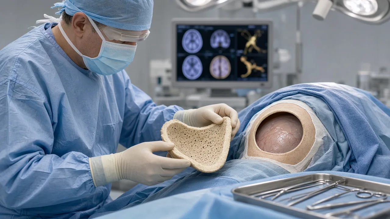
A craniotomy is a type of brain surgery in which the neurosurgeon temporarily removes a section of the skull, called the bone flap, to reach the underlying brain.
Once the required surgical work is completed, the bone flap is normally returned to its original position and secured with specialised plates, screws, wires or other fixation systems.
The procedure itself is therefore an access route to the brain, rather than a treatment for one particular disease.
For example, one patient may require a craniotomy to remove a meningioma, while another may need it to evacuate a haematoma or clip an aneurysm. The surgical technique and risks can be very different in each situation.
Ask whether a craniotomy is appropriate
What Is the Difference Between Craniotomy and Craniectomy?
The terms craniotomy and craniectomy are sometimes used interchangeably by patients, but they are different procedures.
Craniotomy. During a craniotomy, the surgeon temporarily removes a section of skull and generally replaces it at the end of the operation.
Craniectomy. During a craniectomy, the removed portion of skull is not immediately replaced. This may be necessary when the brain is significantly swollen and additional space is needed to reduce pressure. A later operation called a cranioplasty may be performed to reconstruct the skull.
Johns Hopkins also distinguishes burr-hole surgery, which uses a small opening rather than a bone flap. Burr holes are not the same operation as a craniotomy.
The choice depends on the patient's condition and the surgeon's assessment.
Why Is Craniotomy Surgery Performed?
A neurosurgeon may recommend a craniotomy for several different conditions. Not every brain tumour, aneurysm or haematoma requires a craniotomy. Modern neurosurgery also includes minimally invasive procedures, endovascular treatment, stereotactic radiosurgery, medication and observation when clinically appropriate.
1. Brain tumours
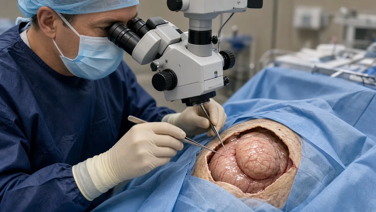
Craniotomy is commonly used to obtain access to brain tumours for biopsy or removal. Depending on the tumour's type, size and location, the goal may be complete removal, maximum safe removal, tissue diagnosis or decompression.
Brain tumours that may require a craniotomy include gliomas, glioblastomas, meningiomas, metastatic brain tumours, oligodendrogliomas, ependymomas, epidermoid tumours, craniopharyngiomas, selected skull-base tumours and other intracranial tumours.
The decision to remove a tumour completely depends on its relationship with important brain structures. In some cases, leaving a small amount of tumour may be safer than attempting complete removal.
Named GAF sheets distinguish glioma surgery from meningioma surgery. Both sit under the brain tumor surgery umbrella when MRI already writes a craniotomy list. Glioma lists sit on Delhi NCR, Mumbai, Bengaluru, Chennai and Hyderabad. Meningioma lists sit on Delhi NCR, Mumbai, Bengaluru, Chennai and Hyderabad.
2. Brain haemorrhage and haematoma
A craniotomy may be required to remove blood or a blood clot that is putting pressure on the brain. This can occur following traumatic brain injury, intracranial haemorrhage, epidural haematoma, subdural haematoma or certain other forms of intracranial bleeding.
Not every brain bleed requires a craniotomy. Treatment depends on the location and size of the haemorrhage, neurological condition and other clinical factors. GAF does not publish a standalone haematoma-craniotomy sheet; those cases are quoted after records review.
3. Brain aneurysm
Some cerebral aneurysms can be treated through an open surgical approach. A craniotomy for aneurysm clipping allows the neurosurgeon to access the affected blood vessel and place a clip across the aneurysm to prevent blood from entering it.
Many aneurysms can also be treated using endovascular aneurysm coiling or flow diversion. The appropriate approach depends on the aneurysm's anatomy and the patient's individual circumstances. Coiling planning is $10,000–$25,000 (typically 3–8 nights), which is a different product from open clipping.
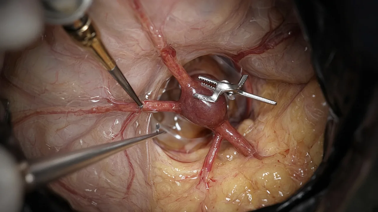
4. Arteriovenous malformation
An arteriovenous malformation (AVM) is an abnormal connection between arteries and veins. Selected AVMs can be surgically removed through a craniotomy on the AVM surgery sheet. Other patients may be treated using AVM embolization, stereotactic radiosurgery or a combination of treatments. The treatment plan is highly individualised.
AVM lists sit on Delhi NCR, Mumbai, Bengaluru, Chennai and Hyderabad.
5. Brain abscess
A brain abscess is an infected collection within the brain. Depending on its location, size and other factors, surgery may be needed to drain or remove the infected material. Antibiotic therapy is also an important part of treatment. NHS patient information on brain-abscess treatment discusses craniotomy and complications in this setting. Abscess surgery is quoted after case review.
6. Traumatic brain injury
Severe head trauma can cause bleeding, swelling or pressure inside the skull. In selected emergencies, neurosurgery may be required to remove a haematoma, repair skull damage or relieve dangerous intracranial pressure. Emergency trauma lists are quoted after case review rather than from a tumour-surgery sheet.
7. Epilepsy surgery
Some patients with medication-resistant epilepsy may be evaluated for surgery. When the seizure-producing area can be safely identified and surgically accessed, a craniotomy may be part of epilepsy surgery. Specialised brain mapping and monitoring may be used to protect important neurological functions.
Share MRI for a neurosurgical opinion
Types of Craniotomy Surgery
The surgical approach is selected according to the location of the brain lesion and the structures that need to be reached.
Conventional craniotomy. A conventional craniotomy uses a larger bone flap to provide adequate surgical exposure. It remains an important approach for many complex neurosurgical procedures.
Keyhole craniotomy. A keyhole approach uses a relatively small opening to reach selected areas of the brain. It may be appropriate for carefully selected tumours and skull-base conditions. Smaller access does not necessarily mean that every patient is suitable for this technique. Selected endoscopic corridors sit on endoscopic brain surgery rather than on a generic keyhole brochure. See Endoscopic Brain Surgery in India. Named pituitary resection sits on Pituitary Tumor Surgery in India. Named CSF diversion sits on Hydrocephalus Surgery in India.
Stereotactic or image-guided craniotomy. Image guidance can combine preoperative imaging with computer-assisted navigation. This helps the surgical team plan and navigate toward the target while identifying important surrounding structures. A tissue sample without a larger opening may sit on stereotactic brain biopsy.
Awake craniotomy. During an awake craniotomy, the patient is awake for part of the operation so the surgical team can assess functions such as speech and movement while operating near important functional brain regions. It is particularly relevant for selected tumours located near areas responsible for language, movement or other critical functions. Awake technique is not a separate GAF price sheet; it is quoted on the named tumour product after case review.
Skull-base craniotomy. Skull-base approaches provide access to difficult areas around the base of the brain. Examples include pterional, orbitozygomatic, supraorbital, retrosigmoid, suboccipital and translabyrinthine approaches. Named skull base surgery planning is $8,000–$22,000 (typically 5–12 nights). Skull-base lists sit on Delhi NCR, Mumbai, Bengaluru, Chennai and Hyderabad.
Who May Need Craniotomy Surgery?
A craniotomy may be considered when a patient's condition requires direct access to the brain or structures within the skull.
The decision may depend on diagnosis, location of the lesion, size of the lesion, symptoms, neurological examination, MRI or CT findings, blood-vessel anatomy, risk of neurological deterioration, response to non-surgical treatment, overall health and previous treatment.
Not every brain tumour, aneurysm or haematoma requires a craniotomy.
Tests Before Craniotomy Surgery
Before planned surgery, the neurosurgical team generally reviews the patient's medical history, neurological examination and imaging.
Depending on the diagnosis, investigations may include:
- MRI brain. MRI provides detailed images of the brain and can help define the location and characteristics of a tumour or other abnormality. Actual imaging files matter more than a written report alone.
- CT scan. CT is particularly useful in emergency settings and for evaluating bleeding, skull fractures and certain structural abnormalities.
- CT angiography or MR angiography. These tests can help visualise cerebral blood vessels when an aneurysm or vascular abnormality is suspected.
- Digital subtraction angiography. In selected vascular cases, angiography may provide detailed information about blood-vessel anatomy.
- Functional mapping. Patients whose lesions are close to important functional areas may undergo specialised testing to identify language, motor or sensory regions.
- Blood tests. Routine laboratory investigations help evaluate blood counts, clotting and general fitness for surgery.
- Pre-anaesthesia evaluation. An anaesthesiologist assesses the patient's medical condition and determines the safest anaesthesia plan.
How to Prepare for Craniotomy Surgery
Preparation varies depending on whether the operation is planned or an emergency.
For planned surgery, patients may be asked to provide a complete list of medications, inform the medical team about blood thinners, report allergies, complete required blood tests and imaging, follow fasting instructions, stop or adjust selected medications only when instructed by the treating team, avoid smoking where possible, arrange transportation and home support after discharge, and discuss expected recovery and rehabilitation requirements.
Patients should never stop blood thinners or other prescribed medicines independently. Johns Hopkins specifically recommends discussing anticoagulants, aspirin and other medications that affect blood clotting with the treating team before surgery.
Request a named-procedure cost estimate
How Is Craniotomy Surgery Performed?
Although the exact operation differs from patient to patient, the general sequence is as follows.
Step 1: Anaesthesia. Most conventional craniotomies are performed under general anaesthesia. Selected procedures may use specialised awake-craniotomy techniques.
Step 2: Positioning. The patient's head is carefully positioned and stabilised to provide safe access to the surgical area.
Step 3: Scalp incision. The neurosurgeon makes an incision at a location selected according to the surgical target.
Step 4: Creating the bone flap. Specialised surgical instruments are used to create openings in the skull and remove the bone flap.
Step 5: Opening the dura. The dura mater, the protective membrane surrounding the brain, is carefully opened.
Step 6: Accessing the target. The neurosurgeon uses microsurgical techniques and, when appropriate, navigation, intraoperative imaging or neurophysiological monitoring.
Step 7: Treating the condition. Depending on the diagnosis, the surgeon may remove a tumour, obtain a biopsy, evacuate a haematoma, clip an aneurysm, remove an AVM, drain an abscess, treat an epileptic focus, relieve pressure or repair damaged structures.
Step 8: Closing the operation. After completing the procedure, the dura and other tissues are closed. The bone flap is generally returned and secured. Finally, the scalp incision is closed with sutures, staples or another appropriate closure method.
How Long Does Craniotomy Surgery Take?
The duration of craniotomy surgery varies substantially. A relatively straightforward procedure may take a few hours, while complex brain tumours, vascular malformations or skull-base procedures can take considerably longer.
Published Indian hospital information commonly places some craniotomy procedures in the 3–5 hour range, but this should not be treated as a fixed duration.
Factors affecting operating time include type of condition, tumour size, tumour location, blood-vessel involvement, surgical approach, need for brain mapping, need for intraoperative imaging, complexity of reconstruction and the patient's medical condition.
What Happens After Craniotomy?
After surgery, patients are usually monitored closely in a recovery area, neurosurgical unit or intensive care setting.
The medical team may repeatedly assess alertness, speech, vision, limb movement, muscle strength, pupillary responses, blood pressure, breathing and neurological function.
Some patients require ICU monitoring, particularly after complex surgery or emergency procedures. An extended ICU stay can substantially increase the total hospital bill.
Early mobilisation and physiotherapy may be introduced according to the patient's condition.
Craniotomy Recovery
Recovery after craniotomy is not the same for every patient. A patient undergoing surgery for a small, accessible lesion may recover differently from someone who has undergone emergency surgery for severe brain trauma.
During the first few days, patients may experience headache, fatigue, facial or scalp swelling, incision discomfort, nausea, temporary weakness, difficulty concentrating and sleep disturbances.
Some patients require rehabilitation if surgery or the underlying disease has affected movement, speech, balance, swallowing or other functions.
Before discharge, the medical team usually provides instructions concerning wound care, medication, activity, bathing, follow-up appointments, driving, warning symptoms and rehabilitation.
Patients should gradually increase activity rather than attempting to return immediately to their previous routine. Johns Hopkins notes that energy and strength can take several weeks to return and that patients should follow their surgeon's instructions regarding heavy lifting and driving.
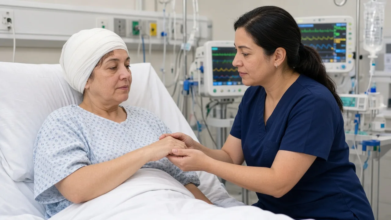
How Long Is the Hospital Stay After Craniotomy?
Hospitalisation varies according to the procedure and the patient's recovery.
GAF planning for brain tumor surgery is typically 5–10 nights. Glioma surgery is typically 5–12 nights. Aneurysm clipping is typically 7–14 nights. Stereotactic brain biopsy is typically 1–3 nights. Johns Hopkins describes a general hospital stay of approximately 3–7 days, although some patients require a longer stay or rehabilitation.
Emergency surgery, neurological deficits, complications or intensive rehabilitation can substantially extend hospitalisation.
Risks and Complications of Craniotomy Surgery
Craniotomy is major brain surgery and carries potential risks. The risks are highly dependent on the location and purpose of the operation.
Possible complications include bleeding, infection, brain swelling, seizures, blood clots, cerebrospinal fluid leakage, anaesthesia-related complications, neurological deficits, speech difficulties, vision problems, weakness or paralysis, memory or cognitive changes, balance or coordination problems, stroke and the need for additional surgery.
The likelihood and severity of these complications vary considerably between patients. For example, surgery close to the brain's speech or motor areas can carry different neurological risks from surgery in another location.
A surgeon should explain the risks that are specifically relevant to the patient's diagnosis and surgical location. UCLH NHS Foundation Trust patient information on craniotomy or biopsy for suspected brain tumour discusses risks and recovery considerations in that setting.
When Should You Contact the Doctor After Craniotomy?
Patients should seek urgent medical advice if they develop symptoms such as increasing or severe headache, new weakness, difficulty speaking, new confusion, seizures, significant changes in vision, fever or chills, increasing redness or swelling around the wound, fluid or blood leaking from the incision, excessive drowsiness, breathing difficulty or chest pain.
These symptoms do not necessarily mean a serious complication has occurred, but they require prompt medical assessment. Sudden severe headache, new paralysis, seizure, chest pain or breathing difficulty belongs in a local emergency department, not in a WhatsApp message.
Craniotomy Surgery Cost in India
The cost of craniotomy surgery in India varies significantly because craniotomy describes a wide range of neurosurgical procedures rather than one standardised operation.
There is no dedicated GAF "craniotomy" package. Planning uses the named product that matches the diagnosis.
| Type / complexity | GAF planning | Typical stay |
|---|---|---|
| Brain tumor surgery | $6,000–$15,000 | 5–10 nights |
| Glioma surgery | $7,000–$16,000 | 5–12 nights |
| Meningioma surgery | $6,500–$15,000 | 5–10 nights |
| Skull base surgery | $8,000–$22,000 | 5–12 nights |
| Endoscopic brain surgery | $5,000–$12,000 | 3–7 nights |
| Stereotactic brain biopsy | $2,000–$6,000 | 1–3 nights |
| Aneurysm clipping | $8,000–$18,000 | 7–14 nights |
| Aneurysm coiling | $10,000–$25,000 | 3–8 nights |
| AVM surgery | $8,000–$20,000 | 7–14 nights |
| Epilepsy surgery | $8,000–$20,000 | 5–10 nights |
| Haematoma, trauma or abscess craniotomy | Quoted after case review | Depends on the case |
These figures are indicative rather than fixed package prices. Published hospital estimates vary by city, hospital, diagnosis and complexity. US comparison for brain tumor surgery is $50,000–$150,000.
Request a named-procedure hospital estimate
WhatsApp +91 90443 46292 for an itemised craniotomy estimate
What Is Included in the Cost of Craniotomy?
A hospital quotation may include some or all of the following: neurosurgeon's fees, anaesthesiologist fees, operating-room charges, hospital room, ICU charges, nursing, medicines, surgical consumables, medical equipment, imaging, pathology, neurophysiological monitoring, blood products when required, and postoperative care.
Inclusions differ between hospitals. For international patients, it is important to request a written treatment estimate based on the patient's medical records rather than relying only on a general internet price.
Factors That Affect Craniotomy Cost in India
Diagnosis. A straightforward haematoma evacuation and a complex skull-base tumour operation have very different resource requirements.
Location of the lesion. Deep-seated or anatomically difficult lesions may require specialised techniques.
Surgical complexity. Longer operations, complex reconstruction and additional procedures increase costs.
Hospital. Large tertiary-care hospitals with advanced neurosurgical infrastructure may have different pricing from smaller facilities.
ICU stay. An extended ICU stay can substantially increase the total hospital bill.
Surgical technology. Navigation, intraoperative imaging, neuromonitoring and specialised equipment may add to the cost.
Pathology. Tumour surgery may require detailed histopathological and molecular testing.
Rehabilitation. Patients who develop or already have neurological deficits may require physiotherapy, occupational therapy or speech therapy.
Craniotomy Surgery Cost in Major Indian Cities
The cost can vary between hospitals and even between individual patients within the same city. Live GAF catalog cities for neurosurgery are Delhi NCR, Mumbai, Bengaluru, Chennai and Hyderabad.
| City | Catalog starting points |
|---|---|
| Delhi NCR | Brain tumours, vascular neurosurgery, skull-base surgery, complex neurosurgery |
| Mumbai | Advanced tertiary neurosurgery and oncology |
| Chennai | Neurosurgery, neuro-oncology and complex brain procedures |
| Bengaluru | Neurosurgery, neuro-oncology and minimally invasive approaches |
| Hyderabad | Neurosurgery, neuro-oncology and advanced imaging |
Pune may have tertiary neurological care, but it is not a live catalog filter on this site. A patient's choice should be based on the specific diagnosis and expertise required, rather than city alone.
- Neurosurgery hospitals in Delhi NCR
- Neurosurgery hospitals in Mumbai
- Neurosurgery hospitals in Bengaluru
- Neurosurgery hospitals in Chennai
- Neurosurgery hospitals in Hyderabad
Plan an international craniotomy stay
Why Do International Patients Consider India for Craniotomy?
Patients travelling internationally for neurosurgery generally look for a combination of experienced neurosurgical teams, multidisciplinary hospital infrastructure, advanced diagnostic imaging, neuro-intensive care, neuro-anaesthesia, rehabilitation, access to oncology when required, availability of minimally invasive and image-guided techniques, international-patient coordination and transparent treatment planning.
For complex neurological conditions, multidisciplinary care can be particularly important because treatment may involve neurosurgery, neurology, neuroradiology, oncology, radiation oncology, pathology and rehabilitation.
How to Choose a Neurosurgeon for Craniotomy in India
Choosing a neurosurgeon should be based on the specific condition, not simply on general experience.
Patients and families can ask how frequently the surgeon performs this type of craniotomy, whether the surgeon routinely treats this particular diagnosis, where the lesion is located, what the goal of surgery is, whether complete removal is realistic or maximum safe removal is the objective, whether minimally invasive options are available, whether awake craniotomy is appropriate, whether intraoperative navigation or neuromonitoring will be used, what neurological risks are specific to this operation, what happens if surgery cannot safely remove the entire lesion, how long hospital and ICU stay is likely to be, what rehabilitation could be required, what the alternatives to surgery are, what could happen without surgery, and whether the patient should obtain a second opinion.
Catalog hospitals currently affiliated with tagged brain-tumour or aneurysm lists include Fortis Memorial Research Institute and Artemis Hospital in Delhi NCR, Medanta in Gurugram, Gleneagles Hospital in Mumbai, Apollo Hospitals on Bannerghatta Road in Bengaluru, Apollo Hospital and MGM Healthcare in Chennai, and KIMS Hospitals in Secunderabad and Apollo Jubilee Hills in Hyderabad. A catalog tag is not a volume, outcome or ranking claim.
Named clinicians currently tagged to brain-tumour or related lists include Dr. Sandeep Vaishya, Dr. Aditya Gupta and Dr. Varindera Paul Singh in Delhi NCR; Dr. Suresh Sankhla and Dr. Nitin Dange in Mumbai; Dr. Krishna K N in Bengaluru; Dr. M Balamurugan and Dr. V. R. Roopesh Kumar in Chennai; and Dr. Sujit Kumar Vidiyala and Dr. Alok Ranjan in Hyderabad.
WhatsApp +91 90443 46292 to match a neurosurgeon
Craniotomy vs Other Brain Surgery Options
A craniotomy is not automatically the best approach for every intracranial condition.
Stereotactic radiosurgery. Stereotactic radiosurgery uses precisely targeted radiation without conventional surgical opening of the skull. It may be appropriate for selected small tumours or vascular conditions. Named radiation-oncology sheets include stereotactic radiosurgery (SRS) at $8,500–$18,000 (typically 1–5 sessions) and Gamma Knife at $10,500–$22,000 (typically 1 session).
Endovascular treatment. Certain aneurysms and vascular abnormalities can be treated through blood vessels rather than open brain surgery. See aneurysm coiling and AVM embolization.
Stereotactic biopsy. A small tissue sample may sometimes be obtained through a minimally invasive image-guided approach instead of a larger craniotomy. See stereotactic brain biopsy.
Observation. Some lesions may be monitored using periodic imaging when immediate intervention is not necessary.
Medication. Some neurological conditions can be treated medically without surgery.
The appropriate option depends on the diagnosis, imaging, symptoms and overall clinical situation.
Craniotomy for Brain Tumour: What Patients Should Know
When craniotomy is performed for a brain tumour, the objective is not necessarily to remove every visible abnormality at any cost. Modern neuro-oncology emphasises maximum safe resection where appropriate. The surgical team must balance tumour removal with preservation of neurological function.
Depending on the tumour, treatment may also involve histopathology, molecular testing, radiation therapy, chemotherapy, targeted treatment, immunotherapy in selected situations and regular MRI surveillance. Therefore, the craniotomy may be one part of a broader brain-tumour treatment plan.
Request a brain-tumor treatment plan
Awake Craniotomy in India
An awake craniotomy is a specialised technique used for selected brain lesions located near important functional areas.
The patient may be awake during part of the operation so the team can test functions such as speech, naming, reading, movement, sensation and other cognitive functions. This allows the surgeon to identify functional boundaries while operating.
Awake craniotomy is not suitable for every patient. Patient cooperation, lesion characteristics, medical condition and the location of the abnormality all influence whether it is appropriate. NCBI / StatPearls discusses indications and functional brain mapping considerations for awake craniotomy.
Medical Tourism for Craniotomy in India
For an international patient, planning brain surgery involves more than arranging an operation.
A typical medical-tourism pathway may include medical-record review, neurosurgical opinion, treatment plan, cost estimate, travel planning, hospital admission, surgery and hospitalisation, discharge and follow-up, and rehabilitation when required.
Send MRI/CT images, reports, previous treatment records and relevant laboratory results. Actual imaging files are particularly important. A written MRI report alone may not provide enough information for a neurosurgeon to assess surgical feasibility.
For complex neurosurgery, international patients should allow flexibility in their travel schedule because the actual hospital stay and recovery can change according to clinical circumstances.
How Long Should International Patients Stay in India?
The required stay depends on diagnosis, type of craniotomy, postoperative neurological status, hospitalisation period, pathology reporting, need for rehabilitation and follow-up consultation.
GAF planning distinguishes hospital stay from total stay in India. Brain tumor surgery is typically 5–10 nights in hospital. Even after discharge, additional time near the hospital is often needed for wound review, pathology discussion and fitness to fly. Complex brain tumour or vascular cases can require substantially longer stays.
A treating hospital should provide a more specific estimate after reviewing the patient's medical records.
Recovery After Craniotomy: Timeline
There is no single recovery timeline that applies to every craniotomy.
First few days. The focus is on neurological monitoring, pain control, wound care, mobility and prevention of complications.
First few weeks. Fatigue and headaches can persist. Activity is gradually increased according to medical advice.
Several weeks. Many patients gradually resume more normal daily activities, although recovery may take longer after complex surgery.
Longer-term recovery. Patients with neurological deficits may require ongoing rehabilitation. Patients treated for brain tumours may also need oncology follow-up and repeated MRI scans.
Rehabilitation After Craniotomy
Rehabilitation may be recommended when a patient has difficulty with walking, balance, strength, speech, swallowing, coordination, daily activities or cognitive functions.
Depending on the patient's needs, rehabilitation may involve physiotherapy, occupational therapy, speech and language therapy and neuropsychological support. The need for rehabilitation depends largely on the underlying disease and the part of the brain involved.
Frequently Asked Questions About Craniotomy Surgery in India
Is craniotomy a major surgery?
Yes. Craniotomy is a major neurosurgical procedure because it involves opening the skull to access the brain. The complexity and risk vary substantially according to the underlying condition and surgical location.
Is craniotomy painful?
Patients can experience headache and scalp or incision discomfort after surgery. Pain management is provided by the medical team. Headaches may persist for some time in certain patients.
How many hours does craniotomy surgery take?
The duration varies. Some procedures may take around three hours, while complex operations can take substantially longer. The exact duration depends on the diagnosis, location and surgical complexity.
How many days will I stay in the hospital?
GAF brain tumor surgery planning is typically 5–10 nights. Johns Hopkins gives a general estimate of approximately 3–7 days, but emergency or complex cases may require longer hospitalisation.
Can a patient walk after craniotomy?
Many patients are encouraged to mobilise as soon as it is medically safe. However, the ability to walk depends on the patient's neurological condition and the type of surgery.
Can a craniotomy affect speech?
It can, particularly when surgery is performed near brain regions responsible for language. Functional mapping and awake-craniotomy techniques may be used in selected cases to help preserve important functions.
Can a craniotomy cause seizures?
Seizures are a recognised possible complication of brain surgery, although the individual risk varies according to the underlying disease and surgical location.
Is the skull replaced after craniotomy?
Usually, yes. The bone flap is generally replaced and secured at the end of the operation. This differs from a craniectomy, where the bone may deliberately be left out temporarily.
What is the success rate of craniotomy?
There is no single success rate for craniotomy because it is used to treat many different diseases. The outcome depends on the diagnosis, patient's health, lesion location, neurological status, surgical objective and other treatments. Patients should ask their neurosurgeon for outcome data relevant to their specific diagnosis and procedure rather than relying on a generic craniotomy success rate.
Is craniotomy safe?
Craniotomy can be performed safely in appropriate patients, but it carries significant potential risks because the brain is involved. Individual risk should be discussed with the treating neurosurgeon.
How much does craniotomy cost in India?
There is no generic craniotomy package. GAF brain tumor surgery planning is $6,000–$15,000. Named glioma, meningioma, skull-base, clipping, AVM and epilepsy sheets differ. Complex or emergency cases can cost more and are quoted after records review.
Can international patients get craniotomy surgery in India?
Yes. International patients can seek neurosurgical treatment in India through hospitals with international-patient departments. Medical records and brain imaging should generally be reviewed before travel.
Which city in India is best for craniotomy?
There is no single city that is appropriate for every patient. Delhi NCR, Mumbai, Chennai, Bengaluru and Hyderabad have major tertiary-care hospitals offering neurosurgical services on this catalog. Pune may have tertiary care, but it is not a live GAF catalog city. The more useful question is which neurosurgical team and hospital have relevant expertise for the patient's specific condition.
What to Send for a Craniotomy Opinion in India
International patients seeking an initial treatment opinion should ideally provide MRI brain images and report, CT scan and report if available, angiography/CTA/MRA if performed, biopsy or pathology reports, previous operation reports, discharge summaries, current medication list, relevant blood-test results, details of previous radiation or chemotherapy, and a short description of current symptoms.
Actual imaging files are particularly important. A written MRI report alone may not provide enough information for a neurosurgeon to assess surgical feasibility.
Key Takeaways
- Craniotomy is an access procedure used in many types of brain surgery.
- A temporary section of skull is usually removed and replaced after the operation.
- It can be used for brain tumours, bleeding, aneurysms, AVMs, abscesses, trauma and selected epilepsy cases.
- The exact technique depends on the location and nature of the brain condition.
- Awake, image-guided, keyhole and skull-base approaches are available for selected patients.
- Recovery varies considerably and can take several weeks or longer.
- GAF planning uses named sheets: brain tumor surgery $6,000–$15,000, with glioma, meningioma, skull-base, clipping, AVM and epilepsy ranges listed above. Haematoma, trauma and abscess work is quoted after case review.
- Patients should obtain a case-specific written quotation after their medical records and imaging have been reviewed.
- For international patients, treatment planning should include hospitalisation, follow-up, rehabilitation and sufficient recovery time.
Get a Craniotomy Treatment Opinion in India
If you or a family member has been advised to undergo craniotomy surgery, the next step is to have the MRI/CT scans and medical records reviewed by an experienced neurosurgical team.
GAF Healthcare can assist international patients with neurosurgeon consultation, medical-record review, hospital selection, treatment-cost estimates, appointment coordination, international-patient assistance, airport and local arrangements and follow-up coordination.
Share your medical reports and brain imaging before making travel arrangements. A case-specific neurosurgical opinion is more useful than relying on a generic procedure description or online cost estimate.
Share imaging for a craniotomy review
Message a coordinator on WhatsApp
Request a cost review on WhatsApp
Top 10 Medical Sources
- Johns Hopkins Medicine — Craniotomy — procedure, indications, risks, preparation, hospital stay and recovery.
- NCBI Bookshelf / StatPearls — Craniotomy — indications, surgical approaches and clinical considerations.
- UCLH NHS Foundation Trust — Craniotomy or Biopsy for Suspected Brain Tumour — risks, recovery considerations and patient information.
- NHS — Brain abscess — craniotomy and complications in brain-abscess treatment.
- Johns Hopkins Medicine — Burr holes — distinction between burr-hole surgery, craniotomy and craniectomy.
- Cleveland Clinic — Craniectomy — distinction between craniectomy and craniotomy and indications for decompressive surgery.
- NCBI / StatPearls — Awake Craniotomy — indications and functional brain mapping considerations.
- GAF Healthcare brain tumor surgery — partner USD planning range for tumour craniotomy lists.
- GAF Healthcare aneurysm clipping — open vascular craniotomy planning range.
- GAF Healthcare skull base surgery and stereotactic radiosurgery — neighbouring corridor and non-open alternatives.
Last reviewed against the cited sources: September 2026.
Medical Disclaimer
This page is intended for educational and medical-tourism information. It does not replace examination, diagnosis or treatment by a qualified neurosurgeon. The need for craniotomy, surgical approach, risks, prognosis and cost must be determined individually after reviewing the patient's clinical history and imaging.
Treatment Process
- 1
Share medical records
The patient provides MRI or CT files, reports, previous treatment records and a short description of current symptoms.
- 2
Neurosurgical review
A neurosurgeon reviews whether the target is a tumour, aneurysm, AVM, haematoma, abscess, epileptic focus or another intracranial condition.
- 3
Procedure selection
The team names the product — brain tumour surgery, glioma, meningioma, skull-base, clipping, AVM, biopsy or another corridor — rather than a generic craniotomy.
- 4
Medical optimisation
Blood thinners, fitness for anaesthesia and any need for mapping, navigation or an awake technique are addressed before planned surgery.
- 5
Itemized estimate
The hospital quotes the named sheet plus ICU, implants, pathology and rehabilitation, not a brochure skull-opening package.
- 6
Travel to India
The patient allows time for in-person examination, additional imaging, surgery, ICU monitoring and early neurological review.
- 7
Surgery
The bone flap is raised, the target is treated, and the bone is generally replaced and secured at the end of the operation.
- 8
Neurological monitoring
Alertness, speech, limb strength and wound care are checked in a neurosurgical unit or ICU according to the case.
- 9
Rehabilitation
Physiotherapy, occupational therapy or speech therapy is added when the disease or the corridor has affected function.
- 10
Return home
The patient leaves with a written summary covering diagnosis, operation performed, pathology, medicines, warning signs and follow-up imaging.