
Treatment Overview
Hydrocephalus surgery in India is performed to treat an abnormal buildup of cerebrospinal fluid (CSF) inside the brain. Depending on the cause, age of the patient, type of hydrocephalus, and findings on brain imaging, treatment may involve a ventriculoperitoneal (VP) shunt, endoscopic third ventriculostomy (ETV), or another form of CSF diversion.
Hydrocephalus can affect newborns, children, young adults and older adults. It may develop because of congenital abnormalities, brain tumours, infections, bleeding, head injury, or blockage of normal CSF circulation. In older adults, normal-pressure hydrocephalus may present primarily with difficulty walking, cognitive changes and urinary problems.
The named endoscopic corridor sits on Endoscopic Brain Surgery in India. Tumour-related blockage often sits on Brain Tumor Surgery in India. A skull opening, when required for tumour or other intracranial work, sits on Craniotomy Surgery in India. Spinal tumours that coexist with CSF problems sit on Spine Tumor Surgery in India. Selected sellar masses sit on Pituitary Tumor Surgery in India.
GAF Healthcare planning for hydrocephalus surgery is $3,000–$8,000 (typically 3–7 nights). US comparison is $20,000–$50,000. Named ETV is $3,000–$8,000 (typically 2–5 nights). Neighbouring endoscopic brain surgery is $5,000–$12,000 (typically 3–7 nights) when a broader neuroendoscopy list is the product. Brain tumor surgery is $6,000–$15,000 (typically 5–10 nights) when tumour removal is required to restore CSF flow. Neighbouring Chiari surgery is $5,000–$12,000 (typically 4–8 nights) when foramen-magnum decompression already belongs on the same dossier. These are planning ranges from partner hospital cost sheets, not hospital quotations. Programmable-valve upgrades, shunt infection, paediatric ICU, ETV with choroid-plexus cauterization, external ventricular drains and combined tumour-plus-shunt lists are quoted after case review.
International patients comparing neurosurgeons commonly start with Delhi NCR, Mumbai, Bengaluru, Chennai and Hyderabad. Endoscopic lists sit on Delhi NCR, Mumbai, Bengaluru, Chennai and Hyderabad. Partner neurosurgery hospitals in Delhi NCR, Mumbai and Bengaluru, and in Chennai and Hyderabad, are a typical first filter. City cost sheets include Delhi NCR, Mumbai, Bengaluru, Chennai and Hyderabad. Kolkata, Pune, Ahmedabad, Chandigarh and Kochi may have hydrocephalus services, but they are not live GAF catalog cities on this site.
India has specialised neurosurgical centres with access to neuroendoscopy, advanced neuroimaging, paediatric neurosurgery, intensive care and multidisciplinary rehabilitation. The appropriate operation is not the same for every patient. A neurosurgeon generally determines whether a shunt, ETV or another intervention is appropriate after reviewing symptoms, MRI or CT findings and the underlying cause.
Important: Hydrocephalus surgery is not a single standardised operation. A VP shunt for communicating hydrocephalus, ETV for aqueductal stenosis, and CSF diversion after a brain tumour are different products with different risks, recovery and cost. This page is educational and does not replace individual specialist advice.
Hydrocephalus Surgery in India at a Glance
| Factor | What patients should know |
|---|---|
| Condition | Abnormal CSF accumulation in the ventricular system |
| Common procedures | VP shunt, ETV, selected ETV-CPC, temporary EVD |
| Anaesthesia | Usually general anaesthesia |
| GAF hospital stay | Typically 3–7 nights for shunt lists; 2–5 nights for named ETV |
| Cost in India | GAF $3,000–$8,000; named sheets differ |
| Patients | Infants, children and adults |
| Follow-up | Essential after any CSF diversion, especially after a shunt |
| Emergency | Sudden drowsiness, vomiting, seizure or return of shunt symptoms belongs in a local emergency department |
What Is Hydrocephalus?
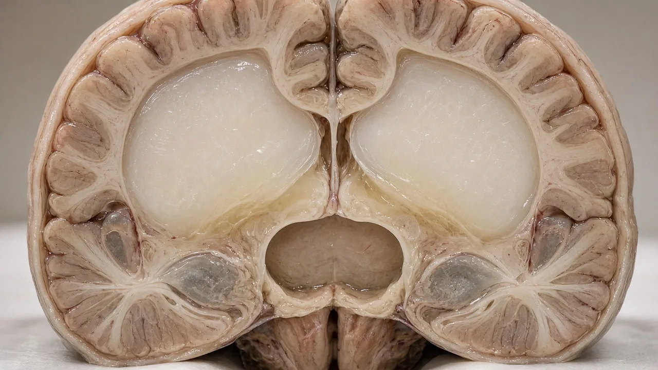
Hydrocephalus is a condition in which cerebrospinal fluid accumulates excessively within the brain's ventricular system.
CSF normally circulates through the ventricles and around the brain and spinal cord before being absorbed into the bloodstream. When production, circulation or absorption becomes abnormal, fluid can accumulate and cause the ventricles to enlarge. Depending on the cause, this enlargement can increase pressure inside the skull and affect brain function.
Hydrocephalus can occur at any age. It is particularly important in infants and young children, but it can also develop later following tumours, infections, bleeding, trauma or other neurological conditions.
Ask whether a shunt or ETV is appropriate
Why Does Hydrocephalus Occur?
There is no single cause. CSF may be unable to flow normally, the body may be unable to absorb it, or brain tissue may have been lost so that the ventricles enlarge without the same mechanism of raised pressure.
Common causes include congenital abnormalities of the brain, aqueductal stenosis, brain tumours, cysts that obstruct CSF flow, meningitis and other infections, intracranial bleeding including subarachnoid haemorrhage, head injury, stroke, complications following neurosurgery, premature birth and intraventricular haemorrhage, myelomeningocele and other congenital conditions, and normal-pressure hydrocephalus in older adults.
The underlying cause matters because it influences whether a patient is considered for ETV, VP shunt surgery, temporary CSF drainage or treatment of the underlying lesion itself.
Types of Hydrocephalus
Obstructive (non-communicating) hydrocephalus occurs when CSF flow is blocked somewhere within the ventricular system. Aqueductal stenosis is one example. Because ETV creates an alternative route for CSF flow, it can be considered in selected patients.
Communicating hydrocephalus occurs when CSF can move through the ventricular system but absorption is impaired. It can follow meningitis, brain haemorrhage, subarachnoid haemorrhage, trauma or other inflammatory conditions. A VP shunt may be required.
Congenital hydrocephalus develops before birth or around the time of birth. Premature infants may also develop hydrocephalus following bleeding into the ventricles.
Acquired hydrocephalus develops later because of another medical condition such as a brain tumour, infection, head trauma, intracranial haemorrhage, stroke or postoperative complications.
Normal-pressure hydrocephalus (NPH) primarily affects older adults. Despite the name, the condition involves enlargement of the brain's ventricles and can produce difficulty walking, cognitive or memory problems, and urinary urgency or incontinence. Not every person with enlarged ventricles has NPH.
Symptoms of Hydrocephalus
Symptoms depend on age, how quickly hydrocephalus develops and the underlying cause.
Infants may show rapid increase in head size, a bulging or tense fontanelle, poor feeding, vomiting, excessive sleepiness, irritability, downward deviation of the eyes, seizures or developmental delay. A rapidly enlarging head or deterioration in an infant requires prompt medical assessment.
Older children may develop headaches, nausea or vomiting, blurred or double vision, problems with balance, difficulty walking, poor school performance, changes in behaviour, excessive sleepiness or seizures.
Adults may experience persistent headaches, nausea or vomiting, visual disturbances, balance problems, difficulty walking, memory or concentration problems, personality or behavioural changes, urinary urgency or incontinence, drowsiness or seizures.
In acute hydrocephalus, symptoms can progress quickly and may represent a medical emergency. That is a local emergency department problem, not a WhatsApp question.
When Is Hydrocephalus Surgery Required?
Not every person with enlarged ventricles automatically needs surgery. The decision depends on symptoms, cause, MRI or CT findings, evidence of progressive ventricular enlargement, intracranial pressure, age, previous shunt surgery, previous infection or bleeding, whether CSF flow is obstructed, and overall neurological condition.
Some people without progressive symptoms may be monitored. When hydrocephalus is causing progressive neurological deterioration or dangerous pressure buildup, surgical CSF diversion is often necessary.
How Is Hydrocephalus Diagnosed?
Diagnosis usually combines history, neurological examination and brain imaging.
MRI can show enlarged ventricles, the location of an obstruction, tumours or cysts, brain malformations, and evidence of previous bleeding or infection.
CT can rapidly demonstrate ventricular enlargement and is particularly useful in emergency situations such as suspected acute hydrocephalus, bleeding or head injury.
Cranial ultrasound can assess the ventricles in babies with an open fontanelle. Prenatal ultrasound can sometimes detect hydrocephalus before birth.
The team may also assess eye movements, strength, reflexes, coordination, balance, walking, memory, cognition and sensation. Additional testing may be required in adults being evaluated for NPH.
Actual imaging files matter more than a written report alone.
Hydrocephalus Surgery Options in India
The surgical procedure is selected according to the type and cause of hydrocephalus.
1. Ventriculoperitoneal shunt surgery
A VP shunt is the most commonly used surgical treatment. The surgeon places a catheter into one of the brain's ventricles. The catheter connects to a valve and tubing that runs underneath the skin to the abdomen, where excess CSF can be absorbed.
Typical steps include anaesthesia, a small opening in the skull, positioning of a ventricular catheter, connection to a valve, subcutaneous tunnelling of tubing, placement of the distal end in the abdominal cavity, and closure. The exact shunt system and valve type vary according to the patient's requirements.

2. Endoscopic third ventriculostomy
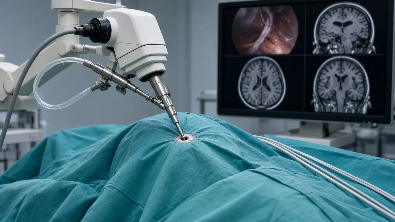
ETV is a minimally invasive procedure that creates an opening in the floor of the third ventricle so CSF can bypass an obstruction. It is particularly associated with obstructive hydrocephalus, including selected aqueductal stenosis.
During ETV the patient is under anaesthesia, a small opening is made in the skull, an endoscope is introduced into the ventricular system, the third ventricle is identified, a small opening is created in its floor, and the incision is closed. Unlike a conventional VP shunt, ETV does not require permanent tubing from the brain to the abdomen. It is not suitable for every patient.
The named ETV product sits on endoscopic third ventriculostomy. The broader corridor product sits on Endoscopic Brain Surgery in India.
3. ETV with choroid plexus cauterization
In selected infants and children, ETV may be combined with choroid plexus cauterization (CPC). The choroid plexus produces CSF. During CPC, selected portions are cauterized with the intention of reducing CSF production.
Suitability varies considerably with age, cause and anatomy. Evidence is particularly heterogeneous in infants and children, so this decision should be made by an experienced paediatric neurosurgical team. There is no separate GAF ETV-CPC cost sheet; these cases are quoted after paediatric records review.
4. External ventricular drain
An EVD temporarily drains CSF from the ventricles into an external collection system. It may be used when rapid control of intracranial pressure is required, such as in selected cases of acute hydrocephalus, brain haemorrhage, severe infection, traumatic brain injury or postoperative complications. It is generally a temporary treatment rather than a definitive long-term solution. There is no separate GAF EVD cost sheet.
Request a named-procedure cost estimate
VP Shunt vs ETV: Which Is Appropriate?
There is no single procedure that is appropriate for every patient.
For paediatric hydrocephalus, evidence-based guidelines recognise both CSF shunts and ETV as treatment options, with the choice influenced by cause, age, anatomy and other clinical factors. ETV can be particularly useful when there is a clear obstruction to CSF flow. A VP shunt may be preferred when ETV is unlikely to provide adequate CSF circulation.
Studies comparing ETV and VP shunting have produced different results depending on the population and cause of hydrocephalus. A 2023 systematic review and meta-analysis of randomised trials in obstructive hydrocephalus found differences in some complication outcomes but no statistically significant difference in operative success, while noting limitations in the available evidence.
The practical question is not simply “Is ETV better than a shunt?” It is: which CSF diversion strategy is most appropriate for this particular patient?
ETV may be considered in aqueductal stenosis, selected tumours causing obstructive hydrocephalus, selected cyst-related obstruction, some cases of recurrent hydrocephalus after shunt surgery, and selected other obstructive conditions. It is generally less suitable when there is widespread impairment of CSF absorption. MRI assessment is an important part of determining whether ETV is technically and clinically appropriate.
Hydrocephalus Surgery in Children
Hydrocephalus in children requires particular attention because the condition can affect brain development. Treatment may involve a VP shunt, ETV, ETV-CPC, temporary ventricular drainage, or treatment of an underlying tumour, cyst or malformation.
Children may require long-term monitoring. A child with a shunt may need further surgery during childhood because of shunt malfunction, infection or growth-related considerations.
Paediatric intensive care, ETV-CPC and combined malformation lists are quoted after case review rather than from the adult $3,000–$8,000 band alone.
Hydrocephalus Surgery in Adults
Adult hydrocephalus has several possible causes, including obstructive hydrocephalus, hydrocephalus following haemorrhage, post-traumatic and post-infectious hydrocephalus, tumour-associated hydrocephalus, normal-pressure hydrocephalus and long-standing ventriculomegaly.
For patients with NPH, careful assessment is particularly important because enlarged ventricles can have other causes. A VP shunt is commonly used when a patient is considered an appropriate surgical candidate. Some investigations may be used to predict whether CSF diversion is likely to improve symptoms.
ETV is not routinely considered the standard treatment for idiopathic NPH. A recent systematic review found that available evidence remains limited and does not support routine ETV for NPH.
Hydrocephalus After Brain Tumor or Hemorrhage
Brain tumours can cause hydrocephalus by blocking normal CSF circulation. Depending on the tumour and location, the team may consider tumour removal, a VP shunt, ETV, temporary ventricular drainage, or a combination. Named tumour resection sits on Brain Tumor Surgery in India.
Bleeding into or around the brain can interfere with CSF circulation and absorption. Treatment may include external ventricular drainage, temporary CSF diversion, a VP shunt, and treatment of the underlying bleeding source. These cases often evolve over time and are quoted after records review.
What Happens Before Hydrocephalus Surgery?
The neurosurgical team usually reviews MRI or CT scans, previous medical records, previous neurosurgical procedures, current symptoms, neurological examination, medications, blood tests, infection risk and anaesthesia requirements.
For international patients travelling to India, it is useful to provide complete imaging files rather than only written reports. A neurosurgeon can make a much more informed assessment when the actual MRI or CT images are available.
A straightforward VP shunt procedure may take approximately one to two hours, although total operating-room time varies. ETV duration varies according to anatomy, cause and complexity. Emergency procedures, revision surgery and cases involving tumours, infection or previous operations may take longer. Use procedure duration as a general guide rather than a guaranteed operating time.
Request a named-procedure hospital estimate
WhatsApp +91 90443 46292 for an itemised hydrocephalus estimate
Hospital Stay and Recovery
GAF planning for hydrocephalus surgery is typically 3–7 nights. Named ETV is typically 2–5 nights. Age, infection, ICU needs, whether the procedure was elective or emergency, and whether additional surgery was performed can lengthen the stay.

Recovery depends heavily on how long the brain has been affected and whether there is permanent neurological injury. Some symptoms may improve relatively quickly after successful CSF diversion. Cognitive, developmental or gait-related problems may take longer.
Children may require physiotherapy, occupational therapy, speech therapy, developmental assessment and educational support. Adults may also benefit from rehabilitation when hydrocephalus has affected walking, cognition or daily activities. Treatment is therefore not always finished when the operation ends.
Patients should avoid planning international travel immediately after surgery without clearance from their treating team.
Risks and Complications
Potential complications include infection, bleeding, seizures, CSF leakage, shunt blockage, shunt disconnection, shunt migration, over-drainage, under-drainage, recurrence of hydrocephalus, ETV closure or failure, neurological injury, and need for revision surgery.
Shunt-related complications are particularly important because a shunt is a mechanical system that can malfunction or become infected. NINDS notes that patients with shunts require monitoring and that some people may require multiple procedures during their lifetime.
Possible warning signs of shunt failure include return of previous hydrocephalus symptoms, headache, nausea or vomiting, increasing sleepiness, vision problems, seizures, fever, redness or tenderness along the shunt pathway, neck or shoulder discomfort, and changes in behaviour or neurological function. A suspected shunt malfunction should be assessed urgently in a local emergency department rather than waiting for the next routine appointment or sending a WhatsApp message.
Can Hydrocephalus Surgery Cure the Condition?
The answer depends on the cause. Hydrocephalus caused by a correctable obstruction may sometimes be treated successfully with ETV or treatment of the underlying cause. Other forms may require long-term CSF diversion. Many patients who undergo shunt surgery continue to require the shunt and lifelong monitoring.
It is more accurate to think of hydrocephalus surgery as controlling abnormal CSF circulation and protecting brain function, rather than assuming that every operation permanently eliminates the underlying condition.
Hydrocephalus Surgery Cost in India
There is no medically responsible single price because treatment may range from a first VP shunt to ETV, programmable-valve revision, paediatric ICU or combined tumour surgery.
| Type / complexity | GAF planning | Typical stay |
|---|---|---|
| Hydrocephalus surgery | $3,000–$8,000 | 3–7 nights |
| ETV | $3,000–$8,000 | 2–5 nights |
| Endoscopic brain surgery | $5,000–$12,000 | 3–7 nights |
| Brain tumor surgery | $6,000–$15,000 | 5–10 nights |
| Chiari surgery | $5,000–$12,000 | 4–8 nights |
| Programmable-valve upgrades, shunt infection, paediatric ICU, ETV-CPC, EVD, combined tumour-plus-shunt | Quoted after case review | Depends on the case |
US comparison for hydrocephalus surgery is $20,000–$50,000.
Major cost factors include procedure type, VP shunt versus ETV, valve and implant type, neurosurgeon fees, hospital category, ICU, length of stay, MRI and CT, laboratory tests, medications, infection treatment, revision surgery, treatment of an underlying tumour, rehabilitation and international-patient services.
A written estimate should clarify specialist consultation, diagnostic work-up, implant, surgeon and operating-room charges, hospital stay, ICU, pathology when relevant, medicines and follow-up. Do not compare two quotations only by the headline price.
City starting points:
- Delhi NCR hydrocephalus surgery cost
- Mumbai hydrocephalus surgery cost
- Bengaluru hydrocephalus surgery cost
- Chennai hydrocephalus surgery cost
- Hyderabad hydrocephalus surgery cost
Plan an international hydrocephalus stay
Why Consider India for Hydrocephalus Surgery?
India has tertiary hospitals with specialist neurosurgery departments, advanced imaging and neurocritical-care infrastructure. A hydrocephalus centre should ideally have adult and paediatric neurosurgery, neuroendoscopy, neuroradiology, neuroanaesthesia, neurocritical care, paediatric intensive care, neurology, rehabilitation, emergency neurosurgical services and infection-control systems that matter for shunt surgery.
Patients should evaluate hospitals based on the named procedure and the complexity of the case, not simply by hospital brand or price. This page does not rank “best hospitals” or claim a national success rate.
Catalog hospitals currently affiliated with tagged hydrocephalus lists include Max Super Speciality Hospital, Saket and Indraprastha Apollo Hospital in Delhi NCR; Gleneagles Hospital and KIMS Hospitals, Thane in Mumbai; Medicover Hospital in Bengaluru; Apollo Hospital and Gleneagles Health City in Chennai; and Yashoda Hospitals, Hi-Tech City and Apollo Hospital, Jubilee Hills in Hyderabad. A catalog tag is not a volume, outcome or ranking claim.
Named clinicians currently tagged to hydrocephalus lists include Dr. Anurag Gupta and Dr. Sogani Shani Kumar in Delhi NCR; Dr. Suresh Sankhla and Dr. Deepu Banerji in Mumbai; Dr. Yashwanth Sandeep in Bengaluru; Dr. M Balamurugan and Dr. Nigel Symss in Chennai; and Dr. M. Vijaya Saradhi and Dr. B G Ratnam in Hyderabad.
Find a hydrocephalus surgeon in India
WhatsApp +91 90443 46292 to match a hydrocephalus surgeon
How to Choose a Hospital for Hydrocephalus Surgery in India
- Neurosurgery hospitals in Delhi NCR
- Neurosurgery hospitals in Mumbai
- Neurosurgery hospitals in Bengaluru
- Neurosurgery hospitals in Chennai
- Neurosurgery hospitals in Hyderabad
For complex hydrocephalus, the experience of the individual neurosurgeon and the broader team can be as important as the hospital name.
What Medical Records Should International Patients Send?
Ideally provide MRI brain images, CT scans if available, radiology reports, previous surgical notes, discharge summaries, blood investigation reports, medication list, previous shunt details, infection history, and neurologist or neurosurgeon consultation notes.
MRI or CT images are generally more useful than sending only the written report, because the neurosurgical team needs to assess the actual anatomy.
Can a Patient Travel to India for Hydrocephalus Surgery?
Yes, for stable patients after records review. Severe or rapidly worsening headache, repeated vomiting, increasing drowsiness, sudden confusion, seizures, new vision problems, sudden difficulty walking, loss of consciousness or rapid neurological deterioration belongs in a local emergency department. For a person with an existing shunt, a sudden return of hydrocephalus symptoms should also be assessed urgently. Do not delay emergency care for international travel planning.
GAF planning distinguishes hospital stay from total stay in India. Hydrocephalus surgery is typically 3–7 nights in hospital. Combined evaluation, imaging, infection watch and fitness to fly usually take longer. Keep the return flight flexible until the treating team confirms fitness for travel.
Request a hydrocephalus treatment plan
Questions to Ask the Neurosurgeon
Ask what is causing the hydrocephalus, whether it is obstructive or communicating, whether there is a tumour, cyst or other blockage, whether MRI shows increased pressure or progressive ventricular enlargement, whether a VP shunt or ETV is more appropriate and why, whether ETV could avoid a permanent shunt in this case, what type of shunt valve will be used, whether a programmable valve is appropriate, which symptoms are expected to improve, how quickly improvement might occur, which neurological problems may not reverse, what happens if the shunt blocks, what the signs of infection are, how frequently revision surgery might be required, how long the patient should remain in India, when the first follow-up is, whether another MRI or CT will be required, and who to contact after returning home.
Frequently Asked Questions About Hydrocephalus Surgery in India
Is hydrocephalus surgery necessary for everyone?
No. Some patients with enlarged ventricles and no progressive symptoms may be monitored. Surgery is generally considered when hydrocephalus is clinically significant, progressive or causing dangerous pressure or neurological impairment.
What is the most common surgery for hydrocephalus?
VP shunt surgery is the most commonly used surgical treatment. ETV is an important alternative for appropriately selected patients, particularly those with obstructive hydrocephalus.
Is ETV better than VP shunt surgery?
Neither procedure is universally better. ETV can be particularly useful when CSF flow is blocked and an internal bypass can be created. VP shunting can be appropriate in many other situations. Clinical evidence comparing the two varies according to age and cause of hydrocephalus.
How much does hydrocephalus surgery cost in India?
GAF hydrocephalus surgery planning is $3,000–$8,000. Named ETV is $3,000–$8,000. A written estimate after records review is required.
How long does hydrocephalus surgery take?
A straightforward VP shunt may take approximately one to two hours, although total operating-room time varies. ETV duration depends on anatomy and cause. Use this as a general guide rather than a guaranteed time.
How many days do I need to stay in the hospital?
GAF planning is typically 3–7 nights after hydrocephalus surgery and 2–5 nights after named ETV. International patients should allow additional time for imaging review and clearance to travel.
Does ETV require a permanent implant?
Usually no. ETV creates an internal opening so it does not require the long catheter system used in a conventional VP shunt. ETV can fail or close, and some patients may subsequently require another procedure or a shunt.
Does a VP shunt stay permanently?
In many patients, yes. Some people continue to need their shunt for life. Children may require revisions as they grow. There is no guaranteed lifespan for a shunt.
Can hydrocephalus come back after surgery?
Yes. Hydrocephalus can recur if a shunt malfunctions or if an ETV closes. Any return of previous symptoms should be evaluated promptly.
Can children live a normal life after shunt surgery?
Many children can participate in school and normal daily activities after successful treatment, although the outcome depends on the cause of hydrocephalus, neurological injury and associated conditions. Some children require ongoing developmental, educational or rehabilitation support.
Can hydrocephalus be treated without surgery?
Some patients with stable ventricular enlargement may be monitored. Established symptomatic hydrocephalus frequently requires surgical CSF diversion. Medicines are generally not a substitute for definitive CSF diversion when there is significant hydrocephalus.
Is ETV suitable for normal-pressure hydrocephalus?
ETV is not routinely considered the standard treatment for idiopathic NPH. Available evidence remains limited and does not support routine ETV for NPH.
Can international patients have hydrocephalus surgery in India?
Yes, subject to medical suitability, travel requirements and the treating hospital's assessment. MRI or CT images should generally be reviewed before travel.
When Should You Seek Urgent Medical Attention?
Go to a local emergency department if a person with known or suspected hydrocephalus develops severe or rapidly worsening headache, repeated vomiting, increasing drowsiness, sudden confusion, seizures, new vision problems, sudden difficulty walking, loss of consciousness or rapid neurological deterioration. For a person with an existing shunt, a sudden return of hydrocephalus symptoms should also be assessed urgently. This is not a WhatsApp question.
Key Takeaways
- Hydrocephalus is caused by abnormal accumulation or circulation of cerebrospinal fluid and can affect infants, children and adults.
- Common causes include congenital abnormalities, tumours, infections, bleeding and trauma.
- VP shunt surgery is the most commonly used surgical treatment. ETV can be an alternative in carefully selected patients, particularly with obstructive hydrocephalus.
- GAF planning for hydrocephalus surgery is $3,000–$8,000 (typically 3–7 nights). Named ETV is $3,000–$8,000 (typically 2–5 nights).
- Treatment should be based on the cause and individual anatomy, not ventricle size alone.
- Shunts can malfunction or become infected and require long-term follow-up.
- Sudden drowsiness, vomiting, seizure or return of shunt symptoms belongs in a local emergency department.
- International patients should share actual MRI or CT images before travelling whenever possible.
Share imaging and previous shunt details before making travel arrangements. A case-specific specialist opinion is more useful than a brochure hydrocephalus package.
Share imaging for a hydrocephalus review
Message a coordinator on WhatsApp
Request a cost review on WhatsApp
Top 10 Medical Sources
- NINDS — Hydrocephalus — causes, diagnosis and treatment, including shunt monitoring.
- Mayo Clinic — Hydrocephalus: symptoms and causes — clinical presentation by age.
- Mayo Clinic — Hydrocephalus: diagnosis and treatment — shunt, ETV and follow-up.
- Hydrocephalus Association — ETV and ETV-CPC — endoscopic alternatives in selected patients.
- Congress of Neurological Surgeons — Pediatric hydrocephalus guidelines — CSF shunt versus ETV.
- Pasqualotto et al., 2023 — systematic review comparing ETV and VP shunt for obstructive hydrocephalus.
- Yadav et al. — Endoscopic third ventriculostomy — indications, technique and complications.
- Sohail et al. — ETV for normal-pressure hydrocephalus — limited evidence against routine ETV for NPH.
- GAF Healthcare hydrocephalus surgery — partner USD planning range for shunt lists.
- GAF Healthcare ETV — neighbouring endoscopic CSF-diversion sheet.
Last reviewed against the cited sources: September 2026.
Medical Disclaimer
This page is intended for general educational purposes and should not replace an examination or treatment recommendation from a qualified neurosurgeon. Hydrocephalus varies substantially in cause, anatomy and previous treatment. The decision to undergo surgery should be based on individual clinical assessment, imaging and the recommendations of the treating team.
If you develop severe or rapidly worsening headache, repeated vomiting, increasing drowsiness, seizures, new vision problems or a sudden return of shunt symptoms, seek urgent medical attention in a local emergency department.
Treatment Process
- 1
Share medical records
The patient provides MRI or CT files, previous shunt details, infection history and a short description of current symptoms.
- 2
Neurosurgical review
A neurosurgeon reviews whether the hydrocephalus is obstructive, communicating, congenital, acquired, tumour-related or NPH.
- 3
Procedure selection
The team names VP shunt, ETV, temporary drainage or tumour treatment rather than a generic hydrocephalus package.
- 4
Medical optimisation
Infection risk, valve type, paediatric ICU needs and anaesthesia fitness are addressed before planned surgery.
- 5
Itemized estimate
The hospital quotes the named sheet plus implant, ICU, imaging and revision risk, not a brochure overnight package.
- 6
Travel to India
Stable patients travel after records review. Sudden drowsiness, vomiting or shunt-failure symptoms are a local emergency.
- 7
Surgery
CSF is diverted through a named shunt or an ETV stoma, with reconstruction or tumour work when those products belong on the list.
- 8
Shunt and neurological monitoring
The team watches for infection, over-drainage, under-drainage, CSF leak and return of hydrocephalus symptoms.
- 9
Rehabilitation and adjuvant planning
Physiotherapy, developmental support or tumour treatment is added when gait, cognition or the underlying cause still need work.
- 10
Return home
The patient leaves with a written summary covering the named procedure, shunt warning signs, imaging schedule and who to contact after returning home.