Treatment Overview
Brain tumor surgery in India is a highly specialised neurosurgical procedure used to diagnose, remove, or reduce a tumour in the brain while protecting as much normal brain function as possible. Depending on the tumour's type, size, location, growth pattern, symptoms, and relationship with critical areas of the brain, surgery may involve complete removal, maximal safe removal, biopsy, or partial tumour removal.
India has advanced neurosurgical centres that provide complex brain tumour treatment using techniques such as microsurgery, neuronavigation, intraoperative monitoring, awake craniotomy, endoscopic surgery, stereotactic biopsy, intraoperative imaging, and minimally invasive approaches. The appropriate technique is not determined by technology alone; it depends on the individual tumour and the patient's neurological condition.
For many patients, surgery is only one part of treatment. Depending on the pathology report and molecular characteristics of the tumour, additional treatment such as radiation therapy, chemotherapy, targeted therapy, or close surveillance may be recommended.
The central principle of modern brain tumour surgery is maximal safe resection: removing as much tumour as can be safely removed while preserving important neurological functions such as speech, movement, vision, memory, swallowing, and coordination.
The skull opening itself is often a craniotomy. This page is the named tumour product. GAF Healthcare planning for brain tumor surgery is $6,000–$15,000 (typically 5–10 nights). Named glioma surgery is $7,000–$16,000 (typically 5–12 nights). Named meningioma surgery is $6,500–$15,000 (typically 5–10 nights). Pituitary tumor surgery is $5,000–$12,000 (typically 3–7 nights). Endoscopic brain surgery is $5,000–$12,000 (typically 3–7 nights). Stereotactic brain biopsy is $2,000–$6,000 (typically 1–3 nights). Skull base surgery is $8,000–$22,000 (typically 5–12 nights). US comparison for brain tumor surgery is $50,000–$150,000. These are planning ranges from partner hospital cost sheets, not hospital quotations.
International patients comparing neurosurgeons commonly start with Delhi NCR, Mumbai, Bengaluru, Chennai and Hyderabad. Glioma lists sit on Delhi NCR, Mumbai, Bengaluru, Chennai and Hyderabad. Partner neurosurgery hospitals in Delhi NCR, Mumbai and Bengaluru, and in Chennai and Hyderabad, are a typical first filter. City cost sheets include Delhi NCR, Mumbai, Bengaluru, Chennai and Hyderabad. Kolkata, Pune, Ahmedabad, Chandigarh and Kochi may have neurosurgical services, but they are not live GAF catalog cities on this site.
Selected brain metastases that sit on a breast or colorectal pathway belong on this named tumour product as well as on Breast Cancer Treatment in India and Colon Cancer Treatment in India. Spinal column disease sits on Spine Tumor Surgery in India.
Important: Brain tumour surgery is not a single standardised operation. The complexity, risks, recovery period and cost depend on the tumour type, location, surgical approach and the patient's neurological condition. This page is educational and does not replace individual neurosurgical advice.
Brain Tumor Surgery in India at a Glance
| Factor | What patients should know |
|---|---|
| Procedure | Biopsy, maximal safe resection or decompression of a brain tumour |
| Common approaches | Craniotomy, awake mapping, endoscopic corridor, stereotactic biopsy |
| Guiding principle | Maximal safe resection, not removal at any cost |
| Hospital stay | GAF brain tumor surgery: typically 5–10 nights |
| Recovery | Weeks to months depending on the tumour and neurological status |
| Cost in India | GAF brain tumor surgery $6,000–$15,000; named sheets differ |
| After surgery | Pathology, molecular tests, radiation or systemic therapy when indicated |
| International patients | Records and imaging review, named-procedure estimate, admission and follow-up |
What Is Brain Tumor Surgery?
Brain tumor surgery refers to a group of neurosurgical procedures used to diagnose or treat tumours arising in the brain or, in some cases, tumours that have spread to the brain from another part of the body.
A tumour may be benign or malignant. Importantly, a benign tumour can still require treatment because its growth may compress or damage important areas of the brain.
Surgery may have several objectives:
- Remove the entire tumour when this can be done safely.
- Remove as much of the tumour as possible.
- Obtain tissue for a precise diagnosis.
- Reduce pressure inside the skull.
- Relieve symptoms caused by the tumour.
- Improve the effectiveness of subsequent radiation or systemic treatment.
- Treat a single symptomatic brain metastasis in selected patients.
For malignant tumours that infiltrate normal brain tissue, surgery generally cannot remove every microscopic tumour cell. In such cases, surgery is often followed by radiation, chemotherapy, targeted treatment, or another appropriate therapy.
Ask whether tumour surgery is appropriate
Why Is Brain Tumor Surgery Needed?
The decision to operate is based on much more than the presence of a tumour on an MRI.
A neurosurgical team considers tumour type, grade, size, exact anatomical location, whether the tumour is superficial or deep, relationship to blood vessels, relationship to speech and movement centres, vision pathways, brainstem involvement, presence of hydrocephalus or increased intracranial pressure, neurological symptoms, the patient's age and general health, molecular characteristics of the tumour, and whether the tumour is newly diagnosed or recurrent.
Some small, slow-growing tumours that cause no symptoms may be monitored rather than immediately operated on. Conversely, a tumour causing significant pressure, seizures, neurological deterioration, hydrocephalus, or other complications may require intervention. Named CSF diversion sits on Hydrocephalus Surgery in India.
Brain Tumor Surgery vs Brain Cancer Surgery
The terms are sometimes used interchangeably, but they are not identical.
A brain tumour may be benign or malignant.
A brain cancer generally refers to a malignant tumour.
Therefore, brain tumour surgery can involve treatment of benign tumours, malignant primary brain tumours, metastatic tumours, tumours of the meninges, skull-base tumours, pituitary-region tumours, certain ventricular tumours, and other intracranial masses requiring tissue diagnosis.
The surgical approach depends on the tumour rather than simply whether it is called "cancer."
Types of Brain Tumor Surgery
There is no single operation called "brain tumor surgery." The procedure is selected according to the anatomy and biology of the tumour.
1. Craniotomy
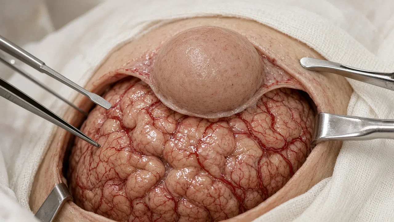
A craniotomy is the most common surgical approach for many brain tumours.
The surgeon makes an incision in the scalp and temporarily removes a section of the skull to gain access to the brain. After the tumour has been treated, the bone flap is generally returned to its original position and secured.
The corridor, risks and recovery of that opening sit on Craniotomy Surgery in India. This page covers the named tumour product that uses that corridor.
Craniotomy may be used for tumour removal, partial tumour removal, biopsy, treatment of selected skull-base tumours, and selected metastatic tumours.
The objective is not simply to remove as much tissue as possible. The objective is to achieve the safest possible tumour removal while protecting normal neurological function.
2. Maximal safe resection
Maximal safe resection is a guiding principle in modern neuro-oncology.
It means removing as much tumour as reasonably possible without causing unacceptable damage to critical neurological functions.
For an infiltrative tumour, attempting to remove every visible abnormality may sometimes increase the risk of permanent neurological disability. A surgeon may therefore intentionally leave a small amount of tumour behind if removing it would threaten speech, movement, vision, memory, or another essential function.
This balance between tumour removal and preservation of neurological function is one of the most important decisions in brain tumour surgery.
3. Awake craniotomy
Awake craniotomy is used for selected tumours located close to areas responsible for important functions such as language or movement.
The patient is appropriately sedated and receives anaesthesia for pain control, but may be awake during part of the procedure. The surgical team can then test specific neurological functions while the surgeon works near critical brain regions.
The purpose is to identify functional areas and help the surgeon remove as much tumour as possible while reducing the risk of neurological injury.
Awake surgery is not appropriate for every patient or every tumour. Patient cooperation, tumour location, medical condition, and the surgical team's assessment all matter. Awake technique is not a separate GAF price sheet; it is quoted on the named tumour product after case review.
4. Stereotactic brain biopsy
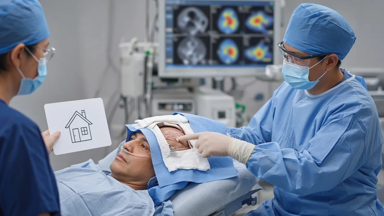
A stereotactic biopsy may be recommended when a tumour is deep-seated, difficult to safely remove, or when obtaining tissue is the primary objective.
Using MRI or CT-based guidance, the neurosurgeon directs a biopsy instrument to the target and removes a small tissue sample. Named GAF stereotactic brain biopsy planning is $2,000–$6,000 (typically 1–3 nights).
The pathologist then examines the tissue to establish the tumour diagnosis and, where appropriate, molecular characteristics that can influence treatment.
A biopsy may therefore be a major part of treatment planning even when tumour removal is not immediately possible.
5. Endoscopic brain surgery
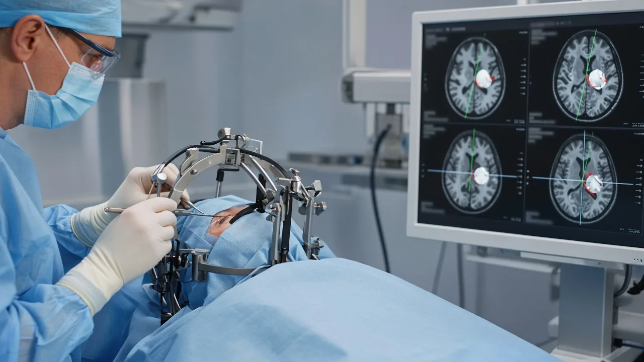
Endoscopic approaches use a thin instrument with a camera to access selected intracranial tumours through a small opening or, for certain skull-base and pituitary tumours, through the nasal passages.
These techniques are particularly relevant for appropriately selected tumours in areas such as the pituitary region and ventricular system. Named endoscopic brain surgery is $5,000–$12,000 (typically 3–7 nights). The named corridor product sits on Endoscopic Brain Surgery in India. Selected sella corridors sit on endoscopic skull base surgery at $6,000–$15,000 (typically 4–8 nights) or on pituitary tumor surgery. Named pituitary resection sits on Pituitary Tumor Surgery in India.
The suitability of endoscopic surgery depends on anatomy and tumour characteristics rather than simply the desire for a smaller incision.
6. Minimally invasive or keyhole brain surgery
Selected tumours can be approached through smaller surgical corridors.
The potential advantages may include smaller incisions and less tissue disruption, but "minimally invasive" does not automatically mean safer or appropriate for every tumour.
The surgical route must provide sufficient access to treat the tumour safely.
7. Intraoperative imaging and navigation-assisted surgery
Modern neurosurgical centres may use image-guided navigation to help the surgeon understand the tumour's three-dimensional position relative to important structures.
Intraoperative imaging can also help account for changes that occur during surgery.
These systems can support surgical precision, but they are tools that complement — not replace — the experience and judgement of the neurosurgical team.
Share MRI for a neurosurgical opinion
Brain Tumors That May Require Surgery
The treatment varies considerably between tumour types.
Surgery may be part of treatment for gliomas, astrocytomas, oligodendrogliomas, glioblastoma, meningiomas, ependymomas, craniopharyngiomas, certain pituitary tumours, schwannomas, selected skull-base tumours, selected metastatic brain tumours, certain paediatric brain tumours, and other intracranial tumours requiring tissue diagnosis.
The exact treatment depends on pathology, molecular findings, location, grade, size, symptoms, and resectability.
Brain Tumor Surgery for Glioblastoma
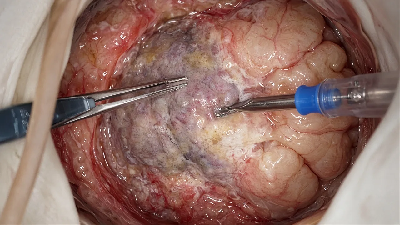
Glioblastoma is an aggressive malignant brain tumour.
For an appropriate surgical candidate, surgery is generally performed to obtain tissue and achieve the greatest safe tumour removal possible. Named glioma surgery planning is $7,000–$16,000 (typically 5–12 nights).
However, surgery alone is generally not sufficient to control glioblastoma because tumour cells can infiltrate surrounding brain tissue beyond what can be seen and safely removed.
Treatment commonly involves surgery followed by radiation therapy ($1,000–$6,000+, typically 15–35 sessions) and chemotherapy ($1,500–$8,000+, outpatient cycles). The NCI identifies surgery followed by radiation and chemotherapy as a standard treatment approach for newly diagnosed glioblastoma.
Brain Tumor Surgery for Meningioma
Meningiomas arise from the meninges, the protective layers surrounding the brain and spinal cord.
Some small, slow-growing and asymptomatic meningiomas may be monitored with regular imaging.
When treatment is required, surgery may be considered depending on the tumour's size, location, growth, symptoms, and relationship with important structures. Named meningioma surgery is $6,500–$15,000 (typically 5–10 nights). Meningioma lists sit on Delhi NCR, Mumbai, Bengaluru, Chennai and Hyderabad.
For selected meningiomas, stereotactic radiosurgery ($8,500–$18,000, typically 1–5 sessions) or Gamma Knife ($10,500–$22,000, typically 1 session) may also be considered, particularly when complete surgical removal is not appropriate or residual disease remains.
Surgery for Brain Metastases
A brain metastasis is cancer that has spread to the brain from another part of the body.
Common primary cancers associated with brain metastases include lung, breast, melanoma, kidney, and colorectal cancers. Selected cases sit beside Breast Cancer Treatment in India and Colon Cancer Treatment in India.
Surgery may be considered for selected patients, particularly when there is a dominant symptomatic lesion causing significant pressure or neurological symptoms and when removal is technically appropriate.
Radiation therapy, stereotactic radiosurgery, systemic therapy, or combinations of these treatments may also be used.
The number of brain lesions, their location, the primary cancer, extracranial disease, overall health, and response to systemic therapy all influence treatment decisions.
How Is a Brain Tumor Diagnosed Before Surgery?
Before surgery, the neurosurgical team needs to understand both the tumour and the surrounding brain.
MRI brain. MRI is one of the most important imaging tests for evaluating brain tumours. Contrast-enhanced MRI may provide detailed information about tumour location, size, enhancement pattern, oedema, and relationship to surrounding structures. Actual imaging files matter more than a written report alone.
CT scan. CT may be useful for evaluating certain tumours, calcification, bleeding, bone involvement, or urgent neurological situations.
Functional MRI. Functional MRI can help identify areas involved in language or movement in selected patients and may contribute to surgical planning.
Diffusion tensor imaging. DTI and tractography can help visualise important white-matter pathways in selected cases.
Angiography. When a tumour is close to important blood vessels or has significant vascularity, vascular imaging may be required.
Biopsy. A biopsy may be performed when tissue diagnosis is needed and complete resection is not appropriate or possible.
Neurological assessment. The team may assess speech, movement, coordination, vision, memory, cognition, and other functions before surgery.
Johns Hopkins notes that MRI, functional MRI, diffusion tensor imaging, CT, and angiography may be used to help plan brain tumour surgery.
What Happens During Brain Tumor Surgery?
Although the exact operation varies, a typical craniotomy-based tumour procedure may follow these stages.
Step 1: Surgical planning. The team reviews MRI and other imaging to determine the safest route to the tumour.
Step 2: Anaesthesia. Most conventional tumour resections are performed under general anaesthesia. Selected awake craniotomy procedures use a carefully planned anaesthesia technique that allows the patient to participate during functional testing.
Step 3: Positioning. The patient's head is positioned and secured to provide stable access to the surgical area.
Step 4: Scalp incision. A carefully planned incision is made according to the surgical approach.
Step 5: Craniotomy. A section of skull is temporarily removed to expose the area of the brain being treated. See Craniotomy Surgery in India.
Step 6: Tumour localisation. The surgeon identifies the tumour using anatomical landmarks, imaging guidance, microscopy, and other available techniques.
Step 7: Tumour removal or biopsy. The surgeon removes the tumour, part of the tumour, or obtains tissue for pathological analysis.
Step 8: Functional monitoring. Depending on the tumour, intraoperative monitoring or brain mapping may be used to help protect important neurological pathways.
Step 9: Closure. The protective layers are closed and the bone flap is generally returned and secured.
Step 10: Recovery monitoring. The patient is transferred to a postoperative recovery area or intensive monitoring unit, depending on the procedure and clinical condition.
Advanced Technologies Used in Brain Tumor Surgery
Technology can improve surgical planning and precision, particularly in complex cases.
Depending on the hospital and clinical indication, available technologies may include a surgical microscope, neuronavigation, intraoperative MRI, intraoperative CT, fluorescence-guided surgery, intraoperative neurophysiological monitoring, functional brain mapping, awake craniotomy, endoscopy, stereotactic navigation, and minimally invasive surgical corridors. Laser-based ablation in selected cases is quoted after records review because GAF does not publish a standalone ablation sheet.
For example, fluorescence-guided surgery can help distinguish certain tumour tissue from surrounding brain tissue in selected tumours, while intraoperative imaging can provide updated anatomical information during an operation.
The availability and usefulness of each technology varies by hospital and by tumour.
Fluorescence-Guided Brain Tumor Surgery
Some brain tumours can be treated using fluorescence-guided techniques.
A fluorescent compound may accumulate preferentially in certain tumour cells and allow them to appear different under specialised surgical illumination. This may help the neurosurgeon identify tumour tissue during resection.
It is important to understand that fluorescence is an aid to surgery, not a guarantee that every tumour cell can be identified or removed.
Brain Mapping and Neurophysiological Monitoring
When a tumour is close to an area responsible for speech, movement, sensation or another critical function, the surgical team may use brain mapping or neurophysiological monitoring.
The objective is to identify and protect important neural pathways during surgery.
This becomes particularly important when the tumour lies within or near what neurosurgeons call eloquent brain regions.
The exact monitoring strategy depends on the tumour location and the patient's neurological functions.
Request a named-procedure cost estimate
How Is the Best Surgical Approach Decided?
There is no universally "best" brain tumour surgery.
The appropriate approach depends on the individual patient: where the tumour is located, how large it is, whether it has a clear boundary, whether it infiltrates normal brain, whether important blood vessels are nearby, whether speech or movement pathways are involved, whether the tumour is superficial or deep, whether the tumour is newly diagnosed or recurrent, the patient's neurological condition, overall health, the expected benefit of surgery, and the risks associated with the proposed surgical route.
This is why two patients with apparently similar "brain tumours" may receive very different treatment plans.
Is Brain Tumor Surgery Always Necessary?
No.
Some tumours can be managed with active surveillance, radiation therapy, stereotactic radiosurgery, chemotherapy, targeted therapy, other systemic treatments, supportive treatment, or a combination of therapies.
Active surveillance may be appropriate for selected slow-growing tumours that do not currently require intervention.
On the other hand, surgery may be particularly important when tissue diagnosis is required, the tumour is safely resectable, significant pressure is being created, or neurological symptoms need to be addressed.
A recommendation against immediate surgery does not necessarily mean that a tumour is being ignored. In selected patients, careful observation is an intentional treatment strategy.
What Happens After Brain Tumor Surgery?
Immediately after surgery, the medical team closely monitors consciousness, speech, limb strength, pupillary responses, vision, vital signs, pain, seizures, brain swelling, possible bleeding, and other neurological changes.
A postoperative MRI or CT scan may be performed to evaluate the surgical site and determine how much tumour remains.
The exact timing of postoperative imaging varies by tumour and clinical circumstances.
Brain Tumor Surgery Recovery
Recovery is highly individual.
Some patients are able to walk shortly after surgery, while others require a longer period of neurological recovery.
GAF planning for brain tumor surgery is typically 5–10 nights. Named glioma surgery is typically 5–12 nights. Stereotactic biopsy is typically 1–3 nights. Complex skull-base or eloquent-area cases can stay longer.
During the early recovery period, patients may experience fatigue, headache, scalp discomfort, temporary swelling, nausea, reduced concentration, weakness, speech difficulties, balance problems, seizures, and emotional changes.
Not every patient experiences these problems, and their severity depends greatly on the tumour and the area treated.
Rehabilitation After Brain Tumor Surgery
Rehabilitation can be an important part of recovery.
Physiotherapy helps improve strength, balance, coordination and mobility.
Speech and language therapy may be required when speech, language or swallowing has been affected.
Occupational therapy helps patients regain independence with everyday activities.
Cognitive rehabilitation may address memory, attention, executive function and other cognitive difficulties.
Psychological and social support can form an important part of recovery for patients and families.
Rehabilitation is not an indication that surgery has failed. It is often part of helping the brain and the patient adapt and recover after treatment.
Risks and Complications of Brain Tumor Surgery
Brain tumour surgery is major surgery, and the risks must be discussed individually with the neurosurgical team.
Potential complications include bleeding, infection, brain swelling, seizures, cerebrospinal fluid leakage, blood clots, stroke, weakness or paralysis, speech or language difficulties, vision changes, balance or coordination problems, memory or cognitive changes, difficulty swallowing, anaesthesia-related complications, need for additional surgery, persistent neurological deficits, and rarely, coma or death.
The risk is strongly influenced by the tumour's location. For example, a tumour close to the motor cortex or language pathways may carry a different neurological risk from a tumour located in a less functionally sensitive region.
Can Brain Tumor Surgery Cause Paralysis?
It can, although the actual risk varies greatly.
The risk depends primarily on whether the tumour is close to or involving pathways responsible for movement.
Modern surgical planning, neuronavigation, functional mapping and intraoperative monitoring may help the surgical team identify and protect important structures in selected cases.
However, no technology can eliminate surgical risk completely.
Patients should ask their neurosurgeon specifically about the estimated risk of weakness or paralysis for their individual tumour rather than relying on a general percentage.
Can Brain Tumor Surgery Affect Speech?
Yes.
Speech and language problems can occur when surgery involves or is close to brain regions responsible for language.
In selected patients, functional MRI, cortical mapping and awake craniotomy may be used to identify language-related areas during surgical planning and treatment.
The purpose is to maximise safe tumour removal while protecting essential functions.
Can a Brain Tumor Come Back After Surgery?
Yes.
Recurrence depends on the tumour's biology, grade, molecular characteristics, location, extent of removal, and additional treatment.
Some benign tumours may recur even after apparently complete removal.
Infiltrative malignant tumours can recur because microscopic tumour cells may remain beyond the visible tumour margin.
Follow-up MRI scans and clinical assessments are therefore an important part of long-term management.
What Happens If the Entire Brain Tumor Cannot Be Removed?
Not being able to remove the entire tumour does not mean treatment has failed.
A surgeon may intentionally leave tumour tissue behind if further removal would create an unacceptable risk to neurological function.
Depending on the pathology, the remaining tumour may then be treated with radiation therapy, stereotactic radiosurgery, chemotherapy, targeted therapy, tumour-specific systemic treatment, observation in selected cases, or clinical trials.
The treatment after surgery is determined by the tumour's pathology and molecular profile.
Brain Tumor Surgery in India
India has developed major tertiary-care centres with dedicated departments for neurosurgery, neuro-oncology, radiation oncology, neuroradiology, neuropathology and rehabilitation.
Live GAF catalog cities are Delhi NCR, Mumbai, Chennai, Bengaluru and Hyderabad. Kolkata, Pune, Ahmedabad, Chandigarh and Kochi may have tertiary neurosurgery, but they are not live catalog filters on this site.
The right hospital should be selected based on the specific tumour rather than city reputation alone.
For a complex brain tumour, patients may benefit from a centre where neurosurgery, neuro-oncology, neuroradiology, neuropathology, radiation oncology, intensive care and rehabilitation work together.
Catalog hospitals currently affiliated with tagged brain-tumour lists include Fortis Memorial Research Institute and Artemis Hospital in Delhi NCR, Gleneagles Hospital in Mumbai, Apollo Hospitals on Bannerghatta Road in Bengaluru, Apollo Hospital and MGM Healthcare in Chennai, and KIMS Hospitals in Secunderabad and Apollo Jubilee Hills in Hyderabad. A catalog tag is not a volume, outcome or ranking claim.
Named clinicians currently tagged to brain-tumour lists include Dr. Sandeep Vaishya, Dr. Aditya Gupta and Dr. Varindera Paul Singh in Delhi NCR; Dr. Suresh Sankhla and Dr. Nitin Dange in Mumbai; Dr. Krishna K N in Bengaluru; Dr. M Balamurugan and Dr. V. R. Roopesh Kumar in Chennai; and Dr. Sujit Kumar Vidiyala and Dr. Alok Ranjan in Hyderabad.
Find a brain tumor neurosurgeon in India
WhatsApp +91 90443 46292 to match a neurosurgeon
Why International Patients Consider India for Brain Tumor Treatment
International patients may consider India because treatment can be coordinated across several specialties within large tertiary hospitals.
For an overseas patient, practical factors can be as important as the operation itself: access to experienced neurosurgical teams, availability of advanced imaging, neuro-ICU facilities, neuro-anaesthesia, neuropathology and molecular testing, radiation oncology, rehabilitation, multidisciplinary tumour boards, international patient services, medical visa support, airport and local transportation, accommodation for family members, and interpreter or language assistance where available.
However, patients should compare the specific clinical capability required for their tumour, rather than choosing a hospital solely because it advertises itself as a leading brain tumour centre.
Cost of Brain Tumor Surgery in India
The cost of brain tumour surgery in India varies substantially from patient to patient.
There is no medically responsible single price for "brain tumor surgery" because the treatment may range from a stereotactic biopsy to a highly complex skull-base or eloquent-area tumour resection.
| Type / complexity | GAF planning | Typical stay |
|---|---|---|
| Brain tumor surgery | $6,000–$15,000 | 5–10 nights |
| Glioma surgery | $7,000–$16,000 | 5–12 nights |
| Meningioma surgery | $6,500–$15,000 | 5–10 nights |
| Pituitary tumor surgery | $5,000–$12,000 | 3–7 nights |
| Endoscopic brain surgery | $5,000–$12,000 | 3–7 nights |
| Stereotactic brain biopsy | $2,000–$6,000 | 1–3 nights |
| Skull base surgery | $8,000–$22,000 | 5–12 nights |
| EBRT after surgery | $1,000–$6,000+ | 15–35 sessions |
| Stereotactic radiosurgery | $8,500–$18,000 | 1–5 sessions |
| Gamma Knife | $10,500–$22,000 | 1 session typical |
| Chemotherapy | $1,500–$8,000+ | Outpatient cycles |
| Targeted therapy | $8,000–$30,000 | Months of therapy |
US comparison for brain tumor surgery is $50,000–$150,000. These figures are indicative rather than fixed package prices.
| Cost factor | Why it matters |
|---|---|
| Tumour type | Different tumours require different treatment pathways |
| Tumour location | Deep or complex tumours may require specialised approaches |
| Tumour size | Larger tumours may require longer or more complex surgery |
| Surgical approach | Craniotomy, endoscopic, stereotactic and other approaches have different costs |
| ICU stay | Longer monitoring increases hospitalisation costs |
| Hospital category | Room and facility charges vary |
| Imaging | MRI, CT and specialised imaging may be required |
| Pathology | Histopathology and molecular testing can add to the cost |
| Navigation/monitoring | Specialised technology may increase procedural costs |
| Radiation or systemic therapy | May be required after surgery for some tumours |
| Rehabilitation | Physiotherapy, speech therapy or other rehabilitation may be needed |
A written estimate should clarify whether the quotation includes surgeon fees, anaesthesia, operating-room charges, ICU, room, medicines, imaging, histopathology, molecular testing, consumables, blood products if required, follow-up consultation and rehabilitation. It is also useful to ask what happens financially if the patient requires a longer ICU stay or an additional procedure.
Request a named-procedure hospital estimate
WhatsApp +91 90443 46292 for an itemised brain tumor estimate
City starting points:
- Delhi NCR brain tumor surgery cost
- Mumbai brain tumor surgery cost
- Bengaluru brain tumor surgery cost
- Chennai brain tumor surgery cost
- Hyderabad brain tumor surgery cost
Plan an international brain tumor stay
How to Choose a Brain Tumor Surgery Hospital in India
Instead of asking only, "Which is the best brain tumor hospital in India?", patients should evaluate the centre against the specific clinical problem.
Important questions include whether the hospital has a dedicated neurosurgery team, neuro-oncology support, advanced imaging, intraoperative monitoring, awake craniotomy when relevant, neuropathology, molecular testing, neuro-ICU care, and rehabilitation.
- Neurosurgery hospitals in Delhi NCR
- Neurosurgery hospitals in Mumbai
- Neurosurgery hospitals in Bengaluru
- Neurosurgery hospitals in Chennai
- Neurosurgery hospitals in Hyderabad
Questions to Ask the Neurosurgeon Before Surgery
Patients and families should consider asking what type of brain tumour is suspected, where exactly it is located, whether it is close to speech, movement, vision or memory areas, whether a biopsy or tumour removal is needed, whether complete removal is realistic, what maximal safe resection means in this case, which neurological functions are most at risk, whether awake craniotomy would be appropriate, whether neuronavigation or intraoperative monitoring will be used, whether tumour tissue will undergo molecular testing, which complications matter most, how long hospital and ICU stay is likely, whether radiation or chemotherapy will be needed after surgery, when the final pathology report will be available, how frequently follow-up MRI is needed, what rehabilitation might be required, when travel and work can resume, and which symptoms after discharge require immediate medical attention.
What Medical Records Should International Patients Send?
Before travelling to India, patients can usually begin by sharing their existing medical records.
Useful documents include latest MRI brain images, MRI report, CT scan and report if available, previous PET/CT where relevant, biopsy report, histopathology report, molecular pathology report, previous operative notes, discharge summaries, current medication list, previous radiation records, previous chemotherapy records, neurological examination reports, and relevant blood investigations.
MRI images themselves are often more useful than only the written MRI report, because the neurosurgeon may need to review the actual images when assessing surgical feasibility.
Patients should retain their original imaging files in DICOM format whenever possible.
Brain Tumor Surgery: Step-by-Step Patient Journey in India
- Send medical records and MRI files.
- A neurosurgeon or multidisciplinary team reviews the case.
- The team determines whether surgery, biopsy, radiation, surveillance or another treatment is appropriate.
- If surgery is recommended, the patient plans travel, admission and accommodation.
- The hospital conducts the required imaging, laboratory tests, anaesthesia evaluation and neurological assessment.
- The planned neurosurgical procedure is performed.
- Tumour tissue is analysed to establish the diagnosis.
- When clinically appropriate, molecular testing further characterises the tumour.
- Postoperative imaging may evaluate residual tumour.
- If additional treatment is needed, neurosurgery, oncology and radiation teams determine the next stage.
- The patient receives postoperative care and rehabilitation where required.
- Long-term clinical and imaging follow-up is planned according to the tumour type.
Request a brain-tumor treatment plan
How Long Should an International Patient Stay in India?
There is no universal duration.
The required stay depends on type of surgery, tumour complexity, recovery, pathology turnaround time, need for molecular testing, need for rehabilitation, and need for radiation or other treatment.
GAF planning distinguishes hospital stay from total stay in India. Brain tumor surgery is typically 5–10 nights in hospital. Even after discharge, additional time near the hospital is often needed for wound review, pathology discussion and fitness to fly. Complex tumours or cases requiring additional treatment can require a considerably longer stay.
International patients should obtain an individualised travel recommendation from the treating hospital rather than booking a fixed return flight immediately after surgery.
When Can a Patient Fly After Brain Tumor Surgery?
Air travel after intracranial surgery should be cleared by the treating neurosurgeon.
The appropriate timing depends on type of surgery, postoperative imaging, presence of brain swelling, seizure risk, CSF leak, wound healing, neurological status, and need for further treatment.
Patients should not assume that a routine commercial flight is safe simply because they feel well.
When Should a Patient Seek Urgent Medical Help After Brain Surgery?
After discharge, urgent medical attention may be required for new or worsening weakness, new difficulty speaking, seizure, loss of consciousness, severe or rapidly worsening headache, repeated vomiting, confusion, significant change in vision, fever with wound problems, fluid leaking from the wound or nose, increasing redness or swelling around the incision, or sudden deterioration in neurological function.
These symptoms belong in a local emergency department, not in a WhatsApp message. The treating hospital should provide individualised postoperative instructions before discharge.
Brain Tumor Surgery in Children
Children with brain tumours require specialised paediatric neurosurgical and neuro-oncology care.
The surgical principles remain similar: establish an accurate diagnosis and remove as much tumour as safely possible while protecting developing brain structures.
However, paediatric surgery has additional considerations involving age and development, tumour biology, blood volume, anaesthesia, neurodevelopment, radiation sensitivity, long-term cognitive effects, rehabilitation, and school and developmental support.
GAF does not publish a standalone paediatric brain-tumour sheet. Children's cases are quoted after paediatric records review rather than from the adult brain tumor surgery band by default.
Brain Tumor Surgery for Older Adults
Age alone does not determine whether brain tumour surgery is possible.
Doctors also consider overall health, functional status, existing neurological deficits, heart and lung function, other medical conditions, tumour characteristics, expected benefit of surgery, and patient preferences.
For some older patients, surgery may provide meaningful symptom relief or diagnostic information. In other situations, less invasive treatment or non-surgical management may be more appropriate.
Brain Tumor Surgery: Myths vs Facts
| Myth | Fact |
|---|---|
| Every brain tumour must be removed completely | Complete removal is not always safe or possible |
| A benign tumour never needs treatment | Benign tumours can cause serious problems through pressure or location |
| Brain surgery always causes permanent neurological problems | Neurological risk varies substantially with tumour location and complexity |
| A biopsy means the tumour cannot be treated | A biopsy can provide essential diagnostic information for selecting treatment |
| A smaller incision always means better surgery | The safest surgical route depends on tumour anatomy and access |
| Brain tumour surgery is always the first step | Some tumours can be observed or treated with radiation or other approaches |
| If part of the tumour remains, surgery has failed | Intentional residual tumour may be appropriate when further removal risks important brain function |
| Recovery is the same for every patient | Recovery varies according to tumour, surgery, neurological status and rehabilitation |
| Brain tumour surgery cures every brain tumour | Some tumours require radiation, chemotherapy, targeted therapy or long-term monitoring after surgery |
Frequently Asked Questions About Brain Tumor Surgery in India
Is brain tumor surgery safe in India?
Brain tumour surgery is performed at specialised neurosurgical centres across India. As with brain surgery anywhere, it carries significant risks. Safety depends on the patient's condition, tumour characteristics, surgical approach, hospital resources and the experience of the treating team.
What is the success rate of brain tumor surgery?
There is no single success rate that applies to all brain tumours. Success may mean obtaining an accurate diagnosis, relieving pressure, achieving maximal safe resection, preserving neurological function, controlling the tumour, or achieving long-term disease control. The expected outcome should therefore be discussed using the specific tumour diagnosis and location.
Can a brain tumor be removed without opening the skull?
Some selected tumours can be approached endoscopically or through minimally invasive techniques. However, many brain tumours still require a craniotomy. The appropriate approach depends on the tumour's location, size and anatomy.
What is maximal safe resection?
Maximal safe resection means removing as much tumour as possible while protecting important neurological functions. It is a central principle in modern brain tumour surgery.
What is awake brain tumor surgery?
Awake craniotomy is a specialised operation in which the patient is awake during part of the procedure so the surgical team can test important neurological functions such as speech or movement. It is used only in appropriately selected patients.
Does every brain tumor need chemotherapy?
No. Chemotherapy is appropriate for certain tumour types and situations. Treatment depends on pathology, molecular characteristics and the overall treatment plan. GAF chemotherapy planning is $1,500–$8,000+.
Does every brain tumor need radiation after surgery?
No. Radiation is recommended for selected tumour types and clinical situations. Some tumours may require surgery alone, while others need radiation, chemotherapy or a combination of treatments. GAF EBRT planning is $1,000–$6,000+.
How long does brain tumor surgery take?
The duration varies according to tumour type, location, size, surgical complexity and the techniques required.
How many days will I stay in the hospital?
GAF brain tumor surgery planning is typically 5–10 nights. Complex cases can require longer hospitalisation. Stereotactic biopsy is typically 1–3 nights.
How long does it take to recover from brain tumor surgery?
Initial recovery may occur over several weeks, while neurological recovery can continue for months. Rehabilitation may be required depending on the patient's symptoms.
Can a brain tumor grow back after surgery?
Yes. The possibility depends on tumour type, grade, molecular characteristics, location and extent of removal.
Is a second surgery possible if the tumor comes back?
In selected patients, repeat surgery may be considered. The decision depends on the location and size of the recurrent tumour, previous treatment, neurological status and overall health.
Can brain tumor surgery affect personality or memory?
It can, particularly when the tumour or surgery involves areas associated with cognition, memory or behaviour. The risk varies substantially according to tumour location.
Can brain tumor surgery affect walking?
It can if the tumour or surgical pathway involves motor areas or movement-related pathways. Preoperative planning and intraoperative monitoring may help reduce this risk in selected cases.
Can brain tumor surgery affect vision?
Yes. The risk depends on the tumour's relationship with the optic nerves, optic pathways and visual processing areas.
Can I get a second opinion before brain tumor surgery?
Yes. For a major neurosurgical decision, obtaining a second opinion can be particularly useful when the tumour is located near a critical brain structure, when surgery has been described as high risk, or when the recommended treatment options differ.
How much does brain tumor surgery cost in India?
GAF brain tumor surgery planning is $6,000–$15,000. Named glioma, meningioma, pituitary, endoscopic, biopsy and skull-base sheets differ. Postoperative radiation and chemotherapy are separate products.
What Makes a Brain Tumor Surgery Center Comprehensive?
For complex cases, patients should look beyond the operating room.
A comprehensive brain tumour programme ideally brings together neurosurgery for surgical planning and tumour removal, neuro-oncology for integrated treatment of malignant brain tumours, neuroradiology for detailed interpretation of MRI, neuropathology for tumour diagnosis and classification, radiation oncology for postoperative or definitive radiation, medical oncology for chemotherapy and targeted therapy, neuro-anaesthesia and neurocritical care, and rehabilitation for recovery of movement, speech, cognition and independence.
A multidisciplinary approach is particularly important when a tumour is complex or requires more than one treatment modality.
Key Takeaways
- Brain tumour surgery is not one single operation; the approach depends on the tumour.
- The primary surgical objective is usually maximal safe tumour removal, not removal at any cost.
- Craniotomy remains a common approach for many brain tumours. The corridor sits on Craniotomy Surgery in India.
- Selected patients may benefit from awake craniotomy, stereotactic biopsy, endoscopic surgery, minimally invasive approaches, neuronavigation or intraoperative monitoring.
- Complete tumour removal is not always possible or safe.
- A biopsy can be essential for determining the exact tumour type and guiding treatment.
- Malignant brain tumours may require radiation, chemotherapy, targeted therapy or other treatment after surgery.
- Recovery varies widely and may include rehabilitation.
- Brain tumour surgery carries important risks, including neurological deficits, bleeding, infection, seizures and stroke.
- GAF planning for brain tumor surgery is $6,000–$15,000 (typically 5–10 nights). Named sheets differ.
- For international patients, the quality of the multidisciplinary team, pathology services, neuro-ICU, rehabilitation and follow-up arrangements are as important as the operation itself.
Share your medical reports and brain imaging before making travel arrangements. A case-specific neurosurgical opinion is more useful than relying on a generic procedure description or online cost estimate.
Share imaging for a tumour review
Message a coordinator on WhatsApp
Request a cost review on WhatsApp
Top 10 Medical Sources
- NCI — Adult Central Nervous System Tumors Treatment (PDQ) — health-professional version covering surgery, radiation and systemic therapy.
- NCI — Adult CNS Tumors Treatment, Patient Version — patient-facing treatment overview.
- American Cancer Society — Surgery for adult brain and spinal cord tumors — surgical goals and recovery considerations.
- Mayo Clinic — Brain tumor — diagnosis and treatment options.
- Mayo Clinic — Craniotomy — skull-opening approach used for many tumours.
- Johns Hopkins Medicine — Brain tumor surgery — imaging, mapping and surgical planning.
- Johns Hopkins Medicine — Brain tumor — treatment pathways including surgery and adjuvant care.
- GAF Healthcare brain tumor surgery — partner USD planning range for tumour resection lists.
- GAF Healthcare glioma surgery and meningioma surgery — named tumour sheets.
- GAF Healthcare stereotactic radiosurgery and Gamma Knife — non-open alternatives for selected lesions.
Last reviewed against the cited sources: September 2026.
Medical Disclaimer
This page is intended for educational purposes and should not replace an examination or treatment recommendation from a qualified neurosurgeon. The appropriate operation, expected benefits, risks, prognosis and treatment cost can only be determined after reviewing the patient's clinical history, imaging and pathology.
Treatment Process
- 1
Share medical records
The patient provides MRI or CT files, reports, previous treatment records and a short description of current symptoms.
- 2
Neurosurgical review
A neurosurgeon reviews whether the target is a glioma, meningioma, pituitary tumour, metastasis or another intracranial mass.
- 3
Procedure selection
The team names resection, maximal safe removal, awake mapping, endoscopic corridor or stereotactic biopsy rather than a generic brain-surgery package.
- 4
Medical optimisation
Fitness for anaesthesia, mapping needs and any adjuvant radiation or systemic therapy are addressed before planned surgery.
- 5
Itemized estimate
The hospital quotes the named sheet plus ICU, pathology, molecular tests and rehabilitation, not a brochure tumour package.
- 6
Travel to India
The patient allows time for in-person examination, additional imaging, surgery, ICU monitoring and pathology.
- 7
Surgery
The tumour is biopsied or removed according to maximal safe resection, then the bone flap is generally replaced.
- 8
Pathology
Histopathology and, when indicated, molecular testing classify the tumour and shape adjuvant treatment.
- 9
Adjuvant planning
Radiation, chemotherapy, targeted therapy, radiosurgery or surveillance is added when the pathology requires it.
- 10
Return home
The patient leaves with a written summary covering diagnosis, residual tumour, medicines, warning signs and MRI follow-up.