
Hearing that a breast examination or mammogram is abnormal can be frightening. An abnormal finding does not automatically mean cancer. Lumps, pain, discharge and appearance changes can come from cysts, fibroadenomas, hormonal shifts or other benign conditions — or they may need further investigation.
Doctors typically use triple assessment: clinical breast examination, breast imaging and pathological examination when indicated. A breast cancer diagnosis is generally confirmed through biopsy, where tissue is examined under a microscope.
This guide covers mammography, ultrasound, MRI, biopsy, BI-RADS, ER/PR/HER2 testing, staging and what international patients should know about diagnosis pathways in India. Companion reading: breast cancer symptoms, diagnosis tests (biopsy & receptors), stages, treatment options and treatment cost in India.
After diagnosis, treatment planning may involve surgery, chemotherapy, radiation, hormone therapy or targeted drugs. GAF planning for lumpectomy is $3,500–$8,000 (typically 1–3 nights) and for mastectomy is $4,500–$10,000 (typically 3–6 nights). Neighbouring sentinel lymph node biopsy is $2,000–$5,500. These are partner planning ranges, not quotations.
Patients comparing surgical oncologists often start with lumpectomy and mastectomy lists alongside medical oncology. Partner hospitals in Delhi NCR, Mumbai and Bengaluru are a typical first filter — see the international patients guide.
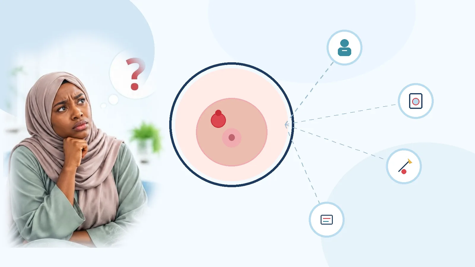
What Is Breast Cancer Diagnosis?
Diagnosis determines whether a breast abnormality is cancerous and characterises the tumour so the team can plan care. Key questions include:
- Is the lump or imaging finding cancerous?
- Is disease invasive or non-invasive (for example DCIS)?
- What histological type and grade are present?
- Are ER, PR and HER2 expressed?
- Are nearby lymph nodes involved?
- Has cancer spread to distant organs?
- Which treatment options fit this biology and stage?
What Is the Triple Assessment for Breast Cancer?
Triple assessment is the standard way to investigate suspicious breast symptoms. The three components complement each other; disagreement between exam, imaging and biopsy prompts further review. National standard treatment guidance in India also emphasises this approach.
| Component | What doctors do | What it helps determine |
|---|---|---|
| Clinical examination | Examine the breast and lymph nodes | Identify suspicious physical changes |
| Breast imaging | Mammography and/or ultrasound (± MRI) | Location and nature of abnormalities |
| Pathological assessment | Examine cells or tissue from biopsy | Confirm or rule out cancer |
Example: a palpable lump that looks suspicious on ultrasound may lead to image-guided core needle biopsy. Relying on a single test increases the chance of missing or misinterpreting disease.
Clinical Breast Examination: The First Diagnostic Step
A breast specialist or trained clinician examines both breasts and regional nodes. They assess size and shape, lump location and feel, skin changes, nipple position or discharge, armpit swelling and nodes near the collarbone. History covers onset, growth, pain, menstrual and reproductive factors, prior breast disease, family history and previous imaging or biopsy.
Examination can flag suspicion but cannot confirm cancer alone. Imaging and tissue testing may still be required — especially when symptoms match patterns described in the symptoms guide.
Mammography: How Does It Detect Breast Cancer?
Mammography uses low-dose X-rays to image breast tissue. It can show small masses, microcalcifications, architectural distortion and other changes that are not obvious on exam.
How is a mammogram performed?
- The patient stands at the mammography unit
- One breast is positioned on the imaging plate
- Gentle compression spreads tissue for a clearer image
- Views are taken from different angles, then repeated for the other breast
Compression may feel briefly uncomfortable and usually lasts only a few minutes. It improves image quality and can reduce the radiation needed.
Screening versus diagnostic mammography
| Feature | Screening mammography | Diagnostic mammography |
|---|---|---|
| Main purpose | Detect cancer before symptoms | Investigate a specific concern |
| Who needs it? | People eligible for routine screening | People with symptoms or abnormal screening |
| Imaging | Standard screening views | Additional targeted views when needed |
| Follow-up | Routine screen or further assessment | May lead to ultrasound or biopsy |
A new breast lump generally needs diagnostic assessment rather than waiting for the next routine screen. Three-dimensional mammography (tomosynthesis) captures multiple angles and may help when overlapping tissue is hard to interpret; availability varies.
Breast Ultrasound: What Does It Show?
Ultrasound uses sound waves to examine palpable lumps or mammographic abnormalities. It helps distinguish fluid-filled cysts from solid masses, map lump location, assess nodes and guide biopsy. The exam is usually painless and uses no ionising radiation.
| Feature | Mammography | Breast ultrasound |
|---|---|---|
| Technology | Low-dose X-rays | Sound waves |
| Microcalcifications | Particularly useful | Limited |
| Cysts vs solid | Sometimes | Very useful |
| Radiation | Low dose | No ionising radiation |
| Common use | Screening and diagnosis | Diagnostic assessment and biopsy guidance |
Ultrasound and mammography are complementary — one does not universally replace the other. Age, breast density, symptoms and findings decide the mix.
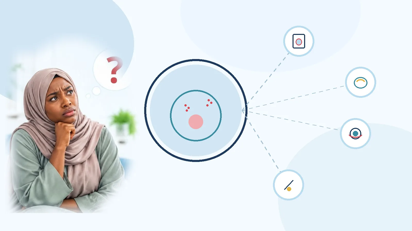
Breast MRI: When Is It Needed?
Breast MRI uses magnetic fields and radio waves, often with gadolinium contrast, for detailed images. It is not required for every lump. Doctors may use it to assess extent of known cancer, evaluate possible multifocal disease or invasive lobular carcinoma, screen selected high-risk women, clarify inconclusive conventional imaging, check response to neoadjuvant therapy or assess some implant cases.
Patients lie face down in a dedicated breast coil; exams often take about 30–60 minutes. MRI is highly sensitive but can also flag non-cancerous findings that need extra work-up — so teams choose it for specific clinical reasons.
What Is a Breast Biopsy?
A biopsy removes a small sample of tissue or cells for laboratory examination. It is generally the definitive way to confirm whether a suspicious area is cancerous. Imaging can look suspicious without proving cancer.
| Biopsy type | How it is performed | Common use |
|---|---|---|
| Fine needle aspiration (FNA) | Thin needle removes cells or fluid | Selected cysts or lymph node assessments |
| Core needle biopsy | Wider needle collects tissue cores | Preferred for most suspicious masses |
| Vacuum-assisted biopsy | Device collects multiple samples | Selected imaging-detected abnormalities |
| Surgical biopsy | Operation removes part or all of an area | When needle biopsy is unsuitable or inconclusive |
Core needle biopsy is commonly preferred because it preserves tissue architecture for type, grade and biomarkers. Image-guided approaches include ultrasound-, stereotactic/mammography- and MRI-guided biopsy when the target cannot be felt.
Is biopsy painful? How long does it take?
Most needle biopsies use local anaesthesia. Patients may feel the anaesthetic injection, pressure, temporary bruising or mild tenderness. A marker clip may be placed for later surgery or imaging. Procedures often take about 15–45 minutes as outpatient care.
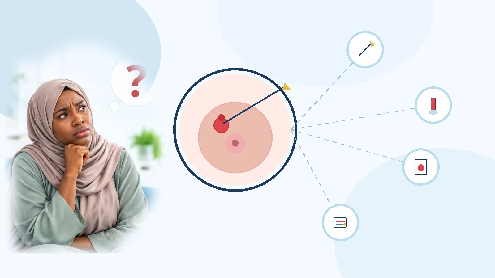
What Happens After a Breast Biopsy?
A pathologist prepares a histopathology report — one of the most important documents in the pathway. It may show a benign finding, a pre-cancerous change or cancer. If cancer is confirmed, the report typically covers type, invasion, grade and receptors. Full walkthrough: pathology report explained.
| Report component | What it means |
|---|---|
| Histological type | Specific type of breast cancer |
| Invasive or non-invasive | Whether cancer has entered surrounding tissue |
| Tumour grade | How abnormal the cells appear |
| DCIS component | Whether ductal carcinoma in situ is present |
| Lymphovascular invasion | Cancer cells in vessels, if identified |
| ER / PR / HER2 | Hormone and HER2 status for treatment selection |
| Ki-67 | Proliferation indicator where tested |
Understanding ER, PR and HER2 Tests
Diagnosis is incomplete without tumour biology. Oestrogen receptor (ER), progesterone receptor (PR) and HER2 status help select hormone therapy, HER2-targeted treatment or other systemic pathways.
| Biomarker | What it identifies | Why it matters |
|---|---|---|
| ER | Oestrogen receptor expression | Hormone therapy eligibility |
| PR | Progesterone receptor expression | Adds hormone-sensitivity context |
| HER2 | Protein overexpression or gene amplification | HER2-targeted treatment options |
| Ki-67 | Tumour cell proliferation | Additional activity information where used |
Triple-negative breast cancer is ER-, PR- and HER2-negative by testing criteria. Chemotherapy (± immunotherapy in eligible cases) often plays a larger role. Deeper detail: ER/PR/HER2 guide and diagnosis tests blog.
Histopathology versus Immunohistochemistry
| Feature | Histopathology | Immunohistochemistry (IHC) |
|---|---|---|
| Main purpose | Examine tissue structure and cancer cells | Identify specific proteins |
| Method | Microscopic tissue examination | Antibody-based staining |
| Confirms cancer type | Yes | Adds biological detail |
| ER/PR/HER2 | Not by morphology alone | Yes (ISH may follow for HER2) |
When HER2 IHC is equivocal, in-situ hybridisation (ISH) may be added. Both histopathology and IHC feed the multidisciplinary plan.
What Is the BI-RADS Classification?
BI-RADS (Breast Imaging Reporting and Data System) standardises how radiologists describe mammography, ultrasound and MRI findings.
| Category | Meaning | Usual next step |
|---|---|---|
| BI-RADS 0 | Incomplete | Additional imaging |
| BI-RADS 1 | Negative | Routine screening |
| BI-RADS 2 | Benign | Routine screening |
| BI-RADS 3 | Probably benign | Short-interval follow-up (often ~6 months) |
| BI-RADS 4 | Suspicious | Biopsy generally recommended |
| BI-RADS 5 | Highly suggestive of malignancy | Tissue diagnosis + specialist assessment |
| BI-RADS 6 | Known biopsy-proven cancer | Treatment planning |
BI-RADS 4 does not mean cancer — it means the finding is suspicious enough for biopsy. BI-RADS 5 is highly suggestive of malignancy, but tissue diagnosis is still required. BI-RADS 3 is probably benign; attend the recommended short-interval follow-up.
How Is Breast Cancer Staged?
After confirmation, staging describes how far disease has spread using the TNM system, often combined with grade and biomarkers for prognostic staging. Overview: breast cancer stages and stages 0–4.
| Component | Meaning | What doctors assess |
|---|---|---|
| T – Tumour | Primary tumour | Size and local extent |
| N – Nodes | Regional lymph nodes | Whether nearby nodes contain cancer |
| M – Metastasis | Distant spread | Whether cancer has reached other organs |
| Stage | General explanation |
|---|---|
| Stage 0 | Non-invasive disease such as DCIS |
| Stage I | Early invasive cancer with limited or no node involvement |
| Stage II | Larger tumour and/or limited regional nodes |
| Stage III | More extensive local or regional disease |
| Stage IV | Distant organ involvement (metastatic) |
Metastatic (Stage IV) disease has spread beyond the breast and regional nodes — commonly to bone, liver, lung or brain. Treatment then focuses on disease control and quality of life alongside biology-specific therapy. See treatment by stage.
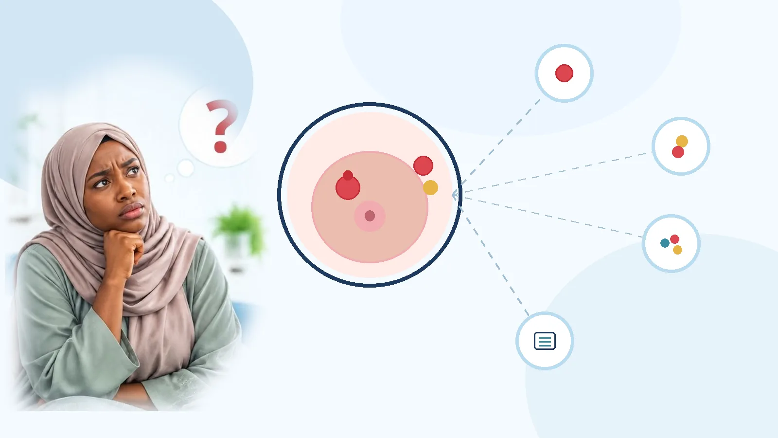
What Tests Check Whether Breast Cancer Has Spread?
Not every patient needs whole-body scanning. Extra tests depend on clinical stage, symptoms and risk.
| Test | Main purpose |
|---|---|
| Abdominal ultrasound | Examine abdominal organs including the liver |
| CT scan | Evaluate organs for suspicious abnormalities |
| PET-CT | Selected cases when advanced disease is suspected |
| Bone scan | Investigate suspected bone involvement |
| Brain MRI | Evaluate suspected brain metastasis |
| Chest imaging | Investigate suspected lung involvement |
| Lymph node biopsy | Confirm suspected nodal involvement |
PET-CT is not compulsory for every early-stage case. Unnecessary scans can find unrelated findings that drive anxiety and extra procedures — teams order staging studies based on need.
What Is Sentinel Lymph Node Biopsy?
The sentinel node is the first drainage node (or group) from the tumour area. Surgeons inject a tracer (radioisotope, blue dye or both), remove the identified node(s) and send them for pathology.
- **Negative sentinel node** — no cancer in sampled node(s)
- **Positive sentinel node** — cancer cells in one or more sampled nodes
A positive node does not automatically mean full axillary clearance; modern decisions are individualised. GAF planning for sentinel lymph node biopsy is $2,000–$5,500 (typically 1–2 nights). Surgery context: breast cancer surgery in India.
How Long Does Breast Cancer Diagnosis Take?
| Diagnostic step | Approximate time |
|---|---|
| Clinical examination | 15–30 minutes |
| Mammography / ultrasound | 15–30 minutes each |
| Breast MRI | 30–60 minutes |
| Needle biopsy | 15–45 minutes |
| Histopathology report | Often several working days |
| IHC / biomarkers | May need additional working days |
| Complete staging | Depends on investigations required |
The full journey may take several days to a few weeks. Ask when each report is due and whether a multidisciplinary board will review results.
What Happens After Breast Cancer Is Confirmed?
Confirmation starts treatment planning. A multidisciplinary team may include surgical, medical and radiation oncologists, radiologists, pathologists, reconstructive surgeons and oncology nurses.
| Factor | Why it matters |
|---|---|
| Cancer stage | Extent of disease |
| Tumour size / nodes | Surgical and systemic choices |
| ER / PR / HER2 | Hormone and targeted options |
| Tumour grade | Behavioural context |
| Menopausal status / health | Treatment suitability |
| Patient preferences | Shared decision-making |
Care may begin with surgery or with medicines first (neoadjuvant therapy / neoadjuvant chemotherapy). Pathways: treatment options, chemotherapy, adjuvant chemotherapy, radiation and reconstruction / reconstruction treatment.
Breast Cancer Diagnosis in India for International Patients
Indian centres offer specialist consultation, mammography, ultrasound, MRI, image-guided biopsy, histopathology and biomarker testing. Patients from East Africa, the Middle East and elsewhere often combine diagnosis review with second opinions and treatment planning.
| Facility | Importance |
|---|---|
| Dedicated breast clinic | Coordinated assessment |
| Experienced breast radiologists | Accurate imaging interpretation |
| Mammography and ultrasound | Essential diagnostic imaging |
| Breast MRI | Selected additional assessment |
| Image-guided biopsy | Accurate tissue sampling |
| Histopathology + IHC | Reliable diagnosis and biomarkers |
| Multidisciplinary tumour board | Integrated treatment planning |
GAF Healthcare can help identify hospitals, coordinate appointments, arrange record review and estimates, and support travel logistics — see international patients.
- Mammography, ultrasound and MRI reports (DICOM images where possible)
- Histopathology, biopsy and ER/PR/HER2 reports
- Prior treatment and surgical records
- Current medications and recent blood tests
Do not delay necessary treatment only to repeat every test abroad. Let the receiving specialist decide what must be redone.
Cost Context After Diagnosis: GAF Planning Ranges
Diagnostic packages vary by hospital, city, imaging mix, biopsy method and pathology panel. GAF does not publish standalone mammography or ultrasound package prices — request a written hospital estimate for the investigations your specialist recommends.
| Investigation area | What influences cost |
|---|---|
| Specialist consultation | Doctor and hospital fees |
| Mammography / ultrasound / MRI | Facility, technique, contrast protocol |
| Core needle biopsy | Guidance method and procedure charges |
| Histopathology + IHC | Tissue processing and biomarker panel |
| Staging CT / PET-CT | Scan type and body coverage when needed |
Once a treatment pathway is clear, use named GAF cost sheets for surgery and systemic care. Full context: breast cancer treatment cost in India.
| Named pathway | GAF planning range in India | Typical stay |
|---|---|---|
| Breast-conserving surgery / lumpectomy | $3,500–$8,000 | 1–3 nights |
| Mastectomy | $4,500–$10,000 | 3–6 nights |
| Oncoplastic breast surgery | $4,500–$11,000 | 2–5 nights |
| Nipple-sparing mastectomy | $5,500–$12,000 | 3–6 nights |
| Breast reconstruction | $6,000–$18,000 | 4–8 nights |
| Sentinel lymph node biopsy | $2,000–$5,500 | 1–2 nights |
| Systemic chemotherapy | $1,500–$8,000+ | Outpatient cycles |
| External beam radiotherapy | $1,000–$6,000+ | Outpatient course |
| Hormone therapy | $1,000–$4,500 | Long-term treatment |
| Targeted therapy | $8,000–$30,000 | Medicine/duration dependent |
| Immunotherapy | $15,000–$45,000 | Repeated outpatient |
| City | Lumpectomy cost sheet | Mastectomy planning |
|---|---|---|
| Delhi NCR | $3,500–$8,000 | $4,500–$10,000 |
| Mumbai | $3,500–$8,000 | $4,500–$10,000 |
| Bengaluru | $3,500–$8,000 | $4,500–$10,000 |
Do not sum every line into one total. Targeted therapy and immunotherapy depend on medicine and duration.
Common Mistakes to Avoid During Diagnosis
- **Assuming every lump is cancer** — many are benign; assess properly
- **Assuming a painless lump is harmless** — cancer is often painless
- **Treating a mammogram as a final diagnosis** — biopsy confirms cancer
- **Ignoring BI-RADS 4 or 5** — follow-up and usually biopsy are needed
- **Starting treatment without adequate pathology** — confirm tissue diagnosis and biomarkers when feasible
- **Ignoring ER/PR/HER2** — these reshape systemic therapy choices
- **Repeating every test unnecessarily** — have specialists review existing reports first
- **Delaying recommended biopsy** — follow advised timelines
Frequently Asked Questions About Breast Cancer Diagnosis
How is breast cancer diagnosed?
Usually through clinical examination, imaging such as mammography and ultrasound, and a biopsy. Tissue examined under a microscope confirms whether cancer is present.
Which test confirms breast cancer?
A breast biopsy with histopathology is generally definitive. Imaging identifies suspicious areas but cannot always confirm cancer.
Is mammography enough to diagnose breast cancer?
No. Mammography detects suspicious changes; ultrasound, MRI and biopsy may still be required.
What is the difference between mammography and breast ultrasound?
Mammography uses low-dose X-rays and is strong for microcalcifications. Ultrasound uses sound waves and helps distinguish cysts from solid masses.
Is breast MRI necessary for every patient?
No. MRI is used selectively — for example high-risk screening, extent assessment or inconclusive conventional imaging.
Does a breast biopsy mean I have cancer?
No. Biopsy investigates an abnormality. Results may be benign, pre-cancerous or cancerous.
Is core needle biopsy better than fine needle aspiration?
Core needle biopsy usually provides tissue architecture and is commonly preferred for suspected breast cancer. FNA has selected uses for cells or fluid.
How long does a breast biopsy report take?
Histopathology often takes several working days. IHC and other tests may add more time depending on the laboratory.
What is BI-RADS 4?
BI-RADS 4 means a suspicious imaging abnormality. Biopsy is generally recommended — it does not by itself confirm cancer.
What does BI-RADS 5 mean?
BI-RADS 5 means imaging is highly suggestive of malignancy. Tissue confirmation is still required.
What does ER-positive or HER2-positive mean?
ER-positive cancers express oestrogen receptors; HER2-positive cancers have increased HER2 expression or amplification. These results guide treatment — see the biomarker guide.
Is PET-CT compulsory for breast cancer staging?
No. PET-CT is selective, especially when advanced disease is suspected. It is not routine for every early-stage case.
Can a blood test diagnose breast cancer?
No routine blood test independently confirms breast cancer. Tissue examination is generally required.
Can breast cancer be missed on a mammogram?
Yes. Persistent symptoms or a suspicious exam may need further imaging or biopsy even after a normal mammogram.
What happens if the biopsy is inconclusive?
Doctors may recommend repeat image-guided biopsy, additional imaging or surgical biopsy after reviewing clinical, imaging and pathology findings together.
Can breast cancer be diagnosed early?
Yes. Screening can find some cancers before symptoms. Early diagnosis helps teams consider treatment while disease is more limited.
Is breast cancer diagnosis available in India?
Yes. Many hospitals offer mammography, ultrasound, MRI, biopsy, histopathology and biomarker testing. Facilities vary — see breast cancer treatment in India.
Should I get a second opinion after diagnosis?
A second opinion can help review biopsy findings, biomarkers, stage and treatment options, especially for complex decisions.
Key Takeaways
- Triple assessment combines exam, imaging and pathology when indicated.
- Biopsy — not imaging alone — generally confirms breast cancer.
- ER, PR and HER2 results are essential for treatment selection.
- BI-RADS 4/5 findings usually need tissue diagnosis.
- Staging uses TNM plus biology; not every patient needs PET-CT.
- GAF surgery planning examples: lumpectomy $3,500–$8,000; mastectomy $4,500–$10,000; SLNB $2,000–$5,500.
- International patients should organise pathology and DICOM imaging before travel.
Top Medical Sources and References
Related Reading on GAF Healthcare
Explore Breast Cancer Care in India
Accurate diagnosis is the foundation of treatment. GAF Healthcare helps international patients coordinate records, specialist consultations and estimates for breast cancer care in India. Website: https://gaf.healthcare/.
Medical Disclaimer
This article is for general educational purposes and does not replace consultation, diagnosis or treatment advice from a qualified professional. Diagnostic tests must be selected by a clinician based on individual findings. Cost figures are indicative planning ranges for named pathways. Emergency symptoms belong in a local emergency department.