
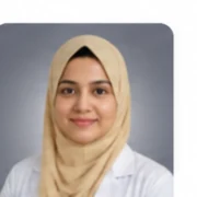
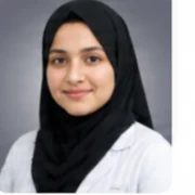
Last updated: 13 September 2026 · Content curated by Dr. Shabnam Choudhary · Medically reviewed by Dr. Saffiyyah Chaudhary
Quick Answer
EBUS (Endobronchial Ultrasound) in Bengaluru is planned against $800–$2,500 per procedure, with Outpatient or 1 night used for broad trip planning. Neither is a local tariff, recommendation or acceptance promise.
Confirm the named interventional pulmonologist trained in linear EBUS, campus, approach, linear ebus mapping and stated needle passes, pathology or microbiology, oxygen plan and where urgent respiratory review would happen.
- India cost range
- $800–$2,500
- Typical starting point
- $800
- Typical hospital stay
- Outpatient or 1 night
- Procedure time
- Often about 45–90 minutes, depending on the number of stations, passes and additional bronchoscopic sampling
- Recovery
- Sore throat, cough, hoarseness, fatigue and small blood streaks may occur briefly.
Major cost factors: number and location of nodal stations, sedation and airway plan, needles and adjunctive devices, rose availability. International patients should also budget for accommodation, airport transfers, a medical visa, medicines and follow-up.
Often quoted separately: changed procedural scope; complications and escalation; premium devices and consumables; advanced diagnostics; travel and ongoing care.
Why request a cost through GAF rather than a hospital?
Writing to one campus gets you that campus’s package. A GAF request is reviewed against your records and returned as suitable doctor and hospital options with an indicative, itemised estimate. There is no obligation to book.
- Doctor review first. The number follows a reading of your imaging, test results and clinical records, not a brochure range.
- Hospital options. You can compare listed campuses before you travel, instead of starting over with each international desk.
- International-patient coordination. Visa letters, records routing and companion logistics sit with the same request.
EBUS (Endobronchial Ultrasound) in Bengaluru
EBUS may be considered when CT or PET shows mediastinal or hilar adenopathy, when invasive nodal staging could change lung-cancer treatment, or when sarcoidosis, tuberculosis, lymphoma or another cause of enlarged nodes is being evaluated. Target accessibility, pre-test probability and whether a larger core or surgical sample is needed should be reviewed first. Planning uses the actual contrast CT and PET images where performed, a map of intended IASLC nodal stations, prior pathology and treatment, oxygen and lung-function assessment, blood count and coagulation review, anticoagulant management and anaesthesia assessment. The team should pre-plan cytology, cell block, microbiology, flow cytometry and molecular tests because one needle rinse cannot reliably serve every question.
The airport is distant from most hospital districts. Name the imaging site, procedure campus and pathology-return visit so cross-city travel is not added during early recovery. Sedation, breathlessness, oxygen and a chest drain can limit independent travel, so arrange a companion.
Accommodation near the confirmed campus is more useful than an airport hotel, especially after sedation or while a chest drain remains. Milder weather does not remove the risk of post-biopsy pneumothorax, fever or oxygen desaturation; complete the scheduled review before onward travel.
Doctor and hospital cards in Bengaluru resolve only from exact live CMS relationships for EBUS (Endobronchial Ultrasound). If that exact relationship is absent, cards must remain empty; a generic Pulmonology or hospital label cannot verify current acceptance. This is a catalog gap, not an availability or quality claim.
Send records before non-refundable travel. A remote opinion can change after examination, image review, oxygen assessment and anaesthesia review in person.
What ebus (endobronchial ultrasound) typically costs in Bengaluru
Systematic multi-station staging takes more scope time, needles and labelled specimens than one targeted aspirate. Moderate sedation differs from general anaesthesia with an endotracheal airway and extended respiratory monitoring.
Ask for clinician, technique, anaesthesia, devices, suite, ward nights, pathology, follow-up and complication terms in writing.
Budget separately for travel through Kempegowda International Airport, nearby lodging, companion support, medicines, oxygen equipment and extra nights if pathology or respiratory monitoring delays departure.
What moves the quote in Bengaluru
- Number and location of nodal stations
- Systematic multi-station staging takes more scope time, needles and labelled specimens than one targeted aspirate.
- Sedation and airway plan
- Moderate sedation differs from general anaesthesia with an endotracheal airway and extended respiratory monitoring.
- Needles and adjunctive devices
- Needle gauge, replacement needles and separately consented forceps or cryobiopsy devices alter consumables.
- ROSE availability
- An on-site cytology team may assess cellularity and redirect passes, but its fee and hours must be stated.
Medical travel through Bengaluru
Send contrast ct chest and pet-ct images and reports with dates, prior bronchoscopy, ebus, pathology, cytology and molecular reports and current oxygen or respiratory-support details where applicable.
Obtain written acceptance from a named interventional pulmonologist trained in linear EBUS and verify the campus, anaesthesia, ICU backup, device availability and pathology pathway.
Sore throat, cough, hoarseness, fatigue and small blood streaks may occur briefly. Patients follow sedation restrictions and await the planned cytology, histology, microbiology and molecular review. Travel waits until oxygen and breathing are stable and the treating team confirms no procedure-related concern. Milder weather does not remove the risk of post-biopsy pneumothorax, fever or oxygen desaturation; complete the scheduled review before onward travel. Carry the procedure, pathology and oxygen or device plan for local follow-up.
Hospitals and units listed in Bengaluru
Doctor and hospital cards in Bengaluru resolve only from exact live CMS relationships for EBUS (Endobronchial Ultrasound). If that exact relationship is absent, cards must remain empty; a generic Pulmonology or hospital label cannot verify current acceptance. This is a catalog gap, not an availability or quality claim.
General accreditation does not establish current EBUS acceptance or equipment. Confirm the named campus has linear EBUS and Doppler equipment, compatible needles, anaesthesia and respiratory rescue capability, and a documented pathway for cytology, cell block, microbiology and molecular specimen handling; ROSE should be claimed only when scheduled.
EBUS (Endobronchial Ultrasound) doctors in Bengaluru · EBUS (Endobronchial Ultrasound) hospitals in Bengaluru · EBUS (Endobronchial Ultrasound) cost in India
What Is EBUS (Endobronchial Ultrasound)?
Endobronchial ultrasound (EBUS) combines flexible bronchoscopy with a linear ultrasound transducer at the scope tip so structures beyond the airway wall can be seen and sampled. EBUS-guided transbronchial needle aspiration (EBUS-TBNA) commonly targets defined mediastinal and hilar lymph-node stations or a central peribronchial mass.

When Might EBUS (Endobronchial Ultrasound) Be Considered?
EBUS may be considered when CT or PET shows mediastinal or hilar adenopathy, when invasive nodal staging could change lung-cancer treatment, or when sarcoidosis, tuberculosis, lymphoma or another cause of enlarged nodes is being evaluated. Target accessibility, pre-test probability and whether a larger core or surgical sample is needed should be reviewed first.
How the operation is performed, recovery and variations →
EBUS (Endobronchial Ultrasound) cost in India
$800–$2,500 is the stored India planning range for EBUS, per procedure, and $4,000–$12,000 the stored self-pay comparison. Neither is a guaranteed package; changed technique, extra pathology, ICU support or a longer stay alters the amount.
A usable estimate names the clinician, campus, approach, linear ebus mapping and stated needle passes, sedation or anaesthesia, oxygen, ward nights, specimen studies and follow-up, with professional, facility, device, imaging and laboratory lines separated.
Budget separately for flights, visa, insurance, transfers, companion, lodging, meals, home oxygen or equipment and local follow-up unless included.
Planning Range ≠ Final Hospital Quotation. Current imaging, respiratory reserve, oxygen needs and assessment by a named interventional pulmonologist trained in linear EBUS come before technique, risks and a final offer are meaningful.
Planning range or quotation?
The $800–$2,500 figure is an indicative planning range. A final hospital quotation is itemised, issued after a consultant reviews your records, and still subject to what is found clinically.
Get a Personalized Cost Estimate
What is usually included in a EBUS (Endobronchial Ultrasound) package?
No two hospitals draw the line in the same place, so read an estimate for what it excludes as carefully as for what it covers. The pattern below is what listed campuses typically bundle into a surgical estimate for this procedure. Anything not written into your estimate should be assumed to be extra until the hospital confirms otherwise.
Usually included
Usually included
Named respiratory consultation
Records and imaging review by the operating pulmonologist when explicitly listed.
Usually included
Pre-procedure assessment
Stated blood tests, ECG, oxygen assessment, urine tests and anaesthesia review; unlisted work-up is extra.
Usually included
Procedure suite and sedation
Bronchoscopy or procedure-room time, standard monitoring, sedation or anaesthesia and recovery care within scope.
Usually included
Quoted hospital stay
The stated day-care or ward category, routine oxygen, nursing and medicines for the listed nights.
Usually included
Standard specimen processing
Histopathology, cytology or microbiology only when itemized; molecular panels and special stains may be separate.
Usually included
Linear EBUS mapping and stated needle passes
The ultrasound bronchoscope, Doppler assessment, specified needles, named node stations and initial specimen preparation only when itemized.
May be charged separately
May be separate
Changed procedural scope
A diagnostic scope becoming therapeutic, rigid-bronchoscopy conversion or an additional biopsy or drainage procedure.
May be separate
Complications and escalation
Unplanned ICU, ventilation, transfusion, chest drain, re-intervention, readmission or extra nights unless covered.
May be separate
Premium devices and consumables
Airway stents, valves, cryoprobes, catheters and single-use scopes beyond the written specification.
May be separate
Advanced diagnostics
PET-CT, molecular or biomarker testing, special microbiology, expert pathology review and repeat imaging unless listed.
May be separate
Travel and ongoing care
Flights, visa, insurance, lodging, companion, home oxygen, rehabilitation and treatment after return.
May be separate
ROSE, advanced assays and repeat tissue acquisition
On-site cytology, flow cytometry, cultures, molecular biomarkers, mediastinoscopy or repeat biopsy are separate unless written.
Catalog inclusions listed for this pathway: pulmonology consultation and records review; named consultant on camera before travel; scope, anaesthesia and overnight stay as quoted; histology, drain or icu step-down as indicated; discharge summary to your home physician.
What can increase the cost?
These are the drivers that actually move a bill for this operation, in rough order of how often they do it. Most of them are clinical decisions rather than commercial ones, which is why an honest estimate is written after a records review rather than before it.
- Number and location of nodal stations
- Systematic multi-station staging takes more scope time, needles and labelled specimens than one targeted aspirate.
- Sedation and airway plan
- Moderate sedation differs from general anaesthesia with an endotracheal airway and extended respiratory monitoring.
- Needles and adjunctive devices
- Needle gauge, replacement needles and separately consented forceps or cryobiopsy devices alter consumables.
- ROSE availability
- An on-site cytology team may assess cellularity and redirect passes, but its fee and hours must be stated.
- Pathology allocation
- Smears, cell block, histology, flow cytometry, special stains and microbiology require separate preparation.
- Cancer biomarkers
- Immunohistochemistry, genomic panels and PD-L1 testing add cost and can still be limited by viable tissue quantity.
- Imaging and physiological testing
- Chest CT or HRCT, PET-CT, spirometry, DLCO, arterial blood gas and echocardiography add lines only when clinically relevant.
- Respiratory reserve and anaesthesia
- Low oxygen, severe airflow limitation, pulmonary hypertension and comorbidity change sedation, monitoring and ICU probability.
- Hospital category and room
- Campus tier, room class, bronchoscopy-suite resources and city shift facility and nursing charges.
- Pathology and follow-up
- Tissue adequacy, microbiology, molecular testing, repeat procedures and review after results alter the complete diagnostic cost.
EBUS (Endobronchial Ultrasound): approaches and where they differ
The approach follows the clinical question, imaging target, airway or pleural anatomy, respiratory reserve and available equipment. Each option has different consumables, monitoring and follow-up, so the quotation must name it.
Swipe to compare surgical approaches →
| Approach | Relative complexity | GAF planning range | Notes |
|---|---|---|---|
| Systematic lung-cancer nodal staging | Individual respiratory assessment determines suitability | No separate GAF sheetRelative complexity only | Accessible mediastinal and hilar stations are mapped and sampled in a sequence designed to avoid contaminating a higher-stage node. Diagnosis, nodal stage and biomarker adequacy are separate endpoints. |
| Targeted EBUS-TBNA | Individual respiratory assessment determines suitability | No separate GAF sheetRelative complexity only | One or more CT- or PET-abnormal nodes or central lesions are sampled to answer a focused diagnostic question, with cytology and cell-block preparation. |
| Granulomatous and infection pathway | Individual respiratory assessment determines suitability | No separate GAF sheetRelative complexity only | Needle material is divided for histology and appropriate tuberculosis, bacterial or fungal studies. Granulomas alone do not establish their cause. |
| EBUS with adjunctive sampling | Individual respiratory assessment determines suitability | No separate GAF sheetRelative complexity only | Standard bronchoscopy, lavage, endobronchial biopsy or selected intranodal forceps/cryobiopsy may be separately consented when aspiration is unlikely to supply enough architecture. |
Planning ranges appear only where GAF Healthcare already publishes a cost sheet for that operation. Other rows describe relative clinical complexity and should not be read as prices.
Diagnostic pulmonology: the subspecialty behind EBUS (Endobronchial Ultrasound)
Diagnostic pulmonology connects respiratory symptoms and chest imaging to physiological tests, airway inspection and tissue or microbiology sampling. The question being answered determines whether a wash, brushing, forceps biopsy, needle aspiration or parenchymal biopsy is appropriate.
Node stations and staging are more than finding an enlarged node
Mediastinal and hilar nodes have named stations tied to anatomic landmarks. In suspected lung cancer, sampling strategy considers the primary tumour, imaging and the highest potential stage first; size or PET uptake alone cannot prove malignant involvement. The report should identify each station and result separately.
EBUS reaches many paratracheal, subcarinal and hilar stations but not every mediastinal compartment. A negative result is interpreted against sample adequacy and disease probability. Surgical staging or another biopsy may remain appropriate when the result would materially alter curative treatment.
Adequacy depends on the diagnostic and biomarker question
ROSE can show lymphocytes, malignant-appearing cells or granulomatous material while the patient is still in the suite, allowing extra passes or another station to be sampled. It is not a substitute for processed cytology, histology, microbiology or expert classification.
Lung-cancer care may require morphology, immunohistochemistry, PD-L1 and genomic testing from the same small sample. Suspected lymphoma may require flow cytometry, while tuberculosis assessment needs sterile material for molecular tests and culture. Pre-labelling containers prevents all material being exhausted by one pathway.
Risks and side effects of EBUS (Endobronchial Ultrasound)
Risks include hypoxaemia, bronchospasm, arrhythmia, sedative reaction, airway bleeding, fever or infection and an inadequate sample. Pneumothorax is uncommon for nodal aspiration but can occur when lung tissue or a peripheral lesion is sampled; rare serious events include major bleeding, mediastinal infection, respiratory failure or injury to an adjacent structure.
Ask before travel who pays for unplanned ventilation, ICU, chest drainage, transfusion, repeat biopsy, readmission or a second procedure.
EBUS (Endobronchial Ultrasound) cost: India vs other medical tourism destinations
India and US values use stored GAF planning ranges; other countries require direct quotations. Compare the same intent, technique, devices, sedation, pathology, ward category and complication terms for EBUS.
Swipe to compare destinations →
| Country | Approximate cost | Relative cost position | Important cost considerations |
|---|---|---|---|
| India | $800–$2,500 | Baseline | GAF catalog planning range. The stored figure covers the named procedure only as written. Diagnostic versus therapeutic scope, anaesthesia, devices, pathology and ICU assumptions must be itemized. |
| Turkey | Confirmation requiredIndicative planning estimate* | Higher than India | Direct quotation required. Compare scope type, biopsy or intervention, anaesthesia, pathology, molecular testing and complication terms rather than a headline package. |
| Thailand | Confirmation requiredIndicative planning estimate* | Higher than India | Direct quotation required. International coordination does not establish tissue adequacy, pathology turnaround, oxygen needs or continuity after return. |
| United Arab Emirates | Confirmation requiredIndicative planning estimate* | Higher than India | Direct quotation required. Pulmonologist, facility, anaesthesia, device, imaging, pathology and follow-up charges may be separate. |
| Singapore | Confirmation requiredIndicative planning estimate* | Higher than India | Private self-pay varies. Request an estimate tied to the imaging target, technique, device, tissue studies and expected admission. |
| Germany | Confirmation requiredIndicative planning estimate* | Higher than India | Private billing varies. Professional billing, eligibility, device scope and postoperative respiratory follow-up require provider confirmation. |
| United Kingdom | Confirmation requiredIndicative planning estimate* | Higher than India | Private self-pay varies. Overseas patients should verify acceptance, urgent respiratory access, pathology delivery and home handover. |
| United States | $4,000–$12,000 | ≈4.8× India | Stored self-pay reference. Pulmonologist, anaesthesia, facility, pathology and device charges may be separate; $4,000–$12,000 is a comparison range, not a quotation. |
International comparisons are indicative. Currency, changed findings, an added device or biopsy, pathology scope and respiratory monitoring alter the final amount.
Why do international patients consider India for EBUS?
Some patients consider India for EBUS because tertiary respiratory teams, interventional bronchoscopy, pleural services, pathology and respiratory critical care can be coordinated with a national planning range. Price alone is not a clinical reason to travel.
Evaluate clinician, licensure, campus, equipment, anaesthesia, ICU backup, pathology quality, urgent access and home handover. No provider is ranked and no outcome is promised; unstable breathing or suitable local care can make travel inappropriate.
EBUS (Endobronchial Ultrasound) hospitals in Bengaluru
Cards follow exact live relationships for EBUS (Endobronchial Ultrasound). A general Pulmonology or accreditation label does not establish current acceptance, equipment or outcomes.
Apollo Hospitals, Bannerghatta Road
 JCI Accredited
 NABH Accredited
- Radiation Oncology
- Surgical Oncology
- Medical Oncology
- Hematology
3 listed doctors for this pathway
Languages listed: English, Kannada, Hindi
Gleneagles Hospitals, Bengaluru
- JCI Accredited
- NABH Accredited
 NABL Accredited
- Radiation Oncology
- Surgical Oncology
- Medical Oncology
- Hematology
1 listed doctor for this pathway
Languages listed: English, Kannada, Hindi
Medicover Hospital, Bengaluru
- JCI Accredited
- NABH Accredited
- Radiation Oncology
- Surgical Oncology
- Medical Oncology
- Hematology
2 listed doctors for this pathway
Languages listed: English, Kannada, Hindi
EBUS (Endobronchial Ultrasound) hospitals in Bengaluru · Talk to a treatment coordinator
EBUS (Endobronchial Ultrasound) specialists in Bengaluru
Profiles appear only when EBUS (Endobronchial Ultrasound) is an exact current CMS relationship. Verify respiratory subspecialty, availability and campus; placement is not a ranking or outcome claim.

Dr. Manjunath B G
Pulmonology
20+ years Experience
Bronchoscopy · EBUS (Endobronchial Ultrasound) · TBNA (Transbronchial Needle Aspiration)
English, Kannada, Hindi
Dr. Manjunath P H
Pulmonology
15+ years Experience
Lung Transplant Surgery · Bronchoscopy · Airway Stenting
English, Kannada, Hindi
Dr. Nanda Kishore K S
Pulmonology
10+ years Experience
Bronchoscopy · EBUS (Endobronchial Ultrasound) · TBNA (Transbronchial Needle Aspiration)
English, Kannada, Hindi
Dr. Ravindra M Mehta
Pulmonology
28+ years Experience
Diagnostic Bronchoscopy · Therapeutic Bronchoscopy · Airway Stenting
English, Kannada, Hindi
Dr. Sudharshan K S
Pulmonology
18+ years Experience
Bronchoscopy · EBUS (Endobronchial Ultrasound) · Medical Thoracoscopy
English, Kannada, Hindi
Dr. Sumant Mantri
Pulmonology
20+ years Experience
Bronchoscopy · EBUS (Endobronchial Ultrasound) · Medical Thoracoscopy
English, Kannada, Hindi
EBUS (Endobronchial Ultrasound) doctors in Bengaluru (6 listed) · Get a personalized cost estimate
EBUS (Endobronchial Ultrasound) cost by city in India
Delhi NCR, Mumbai, Bengaluru, Chennai and Hyderabad use $800–$2,500 because no verified city tariff is stored. City overlays add campus geography, transfers, climate, oxygen logistics and follow-up without inventing prices. Cards require an exact current EBUS (Endobronchial Ultrasound) CMS relationship.
Swipe to compare Indian cities →
Delhi NCR
$800–$2,500
India planning band — not a city quote
Typical stay Outpatient or 1 night
No verified Delhi NCR-only tariff for EBUS is stored. Use $800–$2,500 as the national planning range until a named provider issues an itemized estimate; it is not a city price.
13 hospitals · 40 doctors
Mumbai
$800–$2,500
India planning band — not a city quote
Typical stay Outpatient or 1 night
No verified Mumbai-only tariff for EBUS is stored. Use $800–$2,500 as the national planning range until a named provider issues an itemized estimate; it is not a city price.
4 hospitals · 8 doctors
Bengaluru
$800–$2,500
India planning band — not a city quote
Typical stay Outpatient or 1 night
No verified Bengaluru-only tariff for EBUS is stored. Use $800–$2,500 as the national planning range until a named provider issues an itemized estimate; it is not a city price.
3 hospitals · 6 doctors
Chennai
$800–$2,500
India planning band — not a city quote
Typical stay Outpatient or 1 night
No verified Chennai-only tariff for EBUS is stored. Use $800–$2,500 as the national planning range until a named provider issues an itemized estimate; it is not a city price.
3 hospitals · 8 doctors
Hyderabad
$800–$2,500
India planning band — not a city quote
Typical stay Outpatient or 1 night
No verified Hyderabad-only tariff for EBUS is stored. Use $800–$2,500 as the national planning range until a named provider issues an itemized estimate; it is not a city price.
5 hospitals · 18 doctors
Costs vary considerably by hospital, specialist, clinical complexity, insurance, room or day-care category, and what is included in the package.
Choosing a city for EBUS
What should international patients budget beyond the pulmonology procedure?
International planning starts with records and a named respiratory question. A remote opinion is provisional until examination, image review and anaesthesia assessment confirm the plan.
- Send records
- Provide contrast ct chest and pet-ct images and reports with dates and recent imaging files.
- Remote review
- A named interventional pulmonologist trained in linear EBUS reviews whether EBUS answers the clinical question.
- Define scope
- Name diagnostic or therapeutic intent, technique, samples, devices and likely stay.
- Itemize quotation
- Clinician, suite, anaesthesia, consumables, pathology, ward and exclusions.
- Plan travel
- Flexible flights, accessible lodging, companion and oxygen or equipment logistics.
- Arrive and reassess
- Examination, oxygen assessment, imaging and pre-anaesthetic review.
- Confirm consent
- Purpose, alternatives, risks, possible escalation and sample limitations.
- Complete procedure
- The consented EBUS with respiratory monitoring.
- Early recovery
- Airway, oxygen level, bleeding, pain and sedation recovery.
- Review results
- Imaging, cytology, histopathology or microbiology and any next step.
- Clear travel
- Written travel fitness, medicines, oxygen and urgent-contact plan.
- Handover home
- Procedure note, images, pathology and follow-up schedule for the home team.
- Treatment episode$800–$2,500
- Pre-operative testsOften inside the estimate — confirm
- Hospital stayOutpatient or 1 night typically bundled
- Additional procedures or extended careQuoted separately if advised
- Accommodation for companionVaries by city and length of stay
- Local transportationAirport and daily hospital transfers
- FlightsDepends on origin
- Medical visaFee set by the issuing consulate
Planning estimate — not a hospital quotation.
Get a Personalized Treatment Estimate
What does medical travel for EBUS in India involve?
The sequence below is how a records-first pathway normally runs. The order matters: everything before arrival exists so that you are not making decisions in an unfamiliar hospital corridor with a suitcase beside you.
Define the question
Symptoms, diagnosis, imaging target and prior treatment.
Collect records
CT or HRCT, physiology, pathology and admission history.
Identify specialist
Named interventional pulmonologist trained in linear EBUS and exact campus.
Assess reserve
Oxygen, lung function, comorbidity and anaesthesia fitness.
Choose technique
Diagnostic or therapeutic method for the findings.
Compare quotes
Same scope, devices, samples, nights and follow-up.
Plan travel
Flexible travel, accessible lodging, companion and oxygen.
Confirm consent
Purpose, alternatives, risks and possible escalation.
Complete care
Procedure and monitored recovery.
Process samples
Cytology, histopathology, microbiology or molecular tests.
Attend review
Results, oxygen or device plan and next treatment.
Handover home
Procedure record and respiratory follow-up schedule.

Documents to prepare
- Contrast CT chest and PET-CT images and reports with dates
- Prior bronchoscopy, EBUS, pathology, cytology and molecular reports
- Cancer treatment history or tuberculosis and microbiology records
- Current oxygen needs, lung function, anticoagulants and anaesthetic history
- Recent pulmonology consultation and medication list
- Chest X-ray, CT chest or HRCT reports and image files where available
- Pulmonary function tests including spirometry and DLCO where performed
- Oxygen prescription, CPAP/BiPAP settings and recent admission summaries where relevant
Clinical detail
How the procedure is performed
After topical anaesthetic and sedation or general anaesthesia, the bronchoscope surveys the airway and the linear probe is apposed to the wall. Nodes are identified by station and ultrasound features, vessels are checked with Doppler, and a dedicated needle is advanced through the working channel and airway wall. Multiple passes are placed into labelled containers; rapid on-site evaluation (ROSE), when available, can assess visible cellular material but does not provide a final diagnosis.
Continuous oxygen saturation, rhythm, blood pressure, ventilation and sedation depth are monitored. Recovery includes observation for hypoxaemia, bronchospasm, bleeding, chest pain and anaesthetic effects; fever or infection is assessed clinically. Tissue adequacy, not merely completion of a planned number of passes, determines whether another biopsy route may be discussed.

Main variations
- Systematic lung-cancer nodal staging
- Accessible mediastinal and hilar stations are mapped and sampled in a sequence designed to avoid contaminating a higher-stage node.
- Targeted EBUS-TBNA
- One or more CT- or PET-abnormal nodes or central lesions are sampled to answer a focused diagnostic question, with cytology and cell-block preparation.
- Granulomatous and infection pathway
- Needle material is divided for histology and appropriate tuberculosis, bacterial or fungal studies.
- EBUS with adjunctive sampling
- Standard bronchoscopy, lavage, endobronchial biopsy or selected intranodal forceps/cryobiopsy may be separately consented when aspiration is unlikely to supply enough architecture.
Preparation
Planning uses the actual contrast CT and PET images where performed, a map of intended IASLC nodal stations, prior pathology and treatment, oxygen and lung-function assessment, blood count and coagulation review, anticoagulant management and anaesthesia assessment. The team should pre-plan cytology, cell block, microbiology, flow cytometry and molecular tests because one needle rinse cannot reliably serve every question.
Clinicians direct fasting, inhalers, anticoagulants, diabetes medicines, antibiotics and oxygen. Active infection or unstable breathing may postpone an elective procedure.
Hospital stay and recovery
Uncomplicated EBUS is commonly day care or an overnight observation procedure; limited respiratory reserve, extensive sampling, bleeding, infection concern or anaesthetic recovery can require admission. Sore throat, cough, hoarseness, fatigue and small blood streaks may occur briefly. Patients follow sedation restrictions and await the planned cytology, histology, microbiology and molecular review. Travel waits until oxygen and breathing are stable and the treating team confirms no procedure-related concern.
Follow the oxygen, activity and equipment plan. Seek urgent respiratory help for worsening breathlessness, substantial coughing of blood, persistent or severe chest pain, fever with chills, fainting, confusion or blue lips.
How to compare EBUS (Endobronchial Ultrasound) quotes from Indian hospitals
Print these and work through them on the video call. A house that answers without hedging is telling you something useful about how it will behave when something goes wrong.
- Who is the named interventional pulmonologist trained in linear EBUS, and at which campus?
- What clinical question will EBUS answer or treat?
- Is the scope diagnostic, therapeutic or both?
- Which approach is planned, and what might change it?
- Are pulmonologist, anaesthesia and procedure-suite fees included?
- Which imaging and respiratory tests are needed before the procedure?
- Which scopes, needles, probes, stents, valves, drains or catheters are assumed?
- Which cytology, histopathology and microbiology studies are included?
- Is molecular or biomarker testing included when tissue is obtained?
- How many day-care or ward nights are quoted?
- Are oxygen, non-invasive ventilation and ICU escalation covered?
- What is charged if another procedure or repeat sample is needed?
- How are pneumothorax, bleeding, ventilation and readmission billed?
- When will results be ready, and is the results consultation included?
- Which symptoms require urgent review and where?
- When may I work, exercise and fly?
- Who coordinates respiratory follow-up after I return home?
- Which nodal stations and how many needle passes are planned?
- Is ROSE included and how will material be divided among cell block, microbiology and biomarkers?
- What is the next sampling route if tissue is non-diagnostic or inadequate for molecular testing?
- Are standard bronchoscopy or additional biopsy techniques separately included?
Frequently asked questions
How much does EBUS (Endobronchial Ultrasound) cost in Bengaluru?
$800–$2,500 is the stored national planning range per procedure. No verified Bengaluru-only tariff is stored; request an itemized quotation from a named campus.
Which Bengaluru clinician should assess me for EBUS?
A named interventional pulmonologist trained in linear EBUS should review your records and imaging. Dynamic cards require an exact CMS relationship and are not rankings.
Where should I stay in Bengaluru after EBUS?
Accommodation near the confirmed campus is more useful than an airport hotel, especially after sedation or while a chest drain remains. The airport is distant from most hospital districts. Name the imaging site, procedure campus and pathology-return visit so cross-city travel is not added during early recovery.
How much does EBUS (Endobronchial Ultrasound) cost in India?
EBUS (Endobronchial Ultrasound) is typically planned at $800–$2,500 per procedure. This national range is not a quotation; technique, sedation, devices, pathology, ward nights and written terms determine the amount.
What is EBUS (Endobronchial Ultrasound)?
Endobronchial ultrasound (EBUS) combines flexible bronchoscopy with a linear ultrasound transducer at the scope tip so structures beyond the airway wall can be seen and sampled. EBUS-guided transbronchial needle aspiration (EBUS-TBNA) commonly targets defined mediastinal and hilar lymph-node stations or a central peribronchial mass.
When is EBUS considered?
EBUS may be considered when CT or PET shows mediastinal or hilar adenopathy, when invasive nodal staging could change lung-cancer treatment, or when sarcoidosis, tuberculosis, lymphoma or another cause of enlarged nodes is being evaluated. Target accessibility, pre-test probability and whether a larger core or surgical sample is needed should be reviewed first.
What does the EBUS planning range usually include?
It commonly includes the named respiratory specialist, procedure suite, routine monitoring, sedation or anaesthesia, standard consumables and quoted stay. Imaging, advanced devices, pathology panels and treatment after results may be separate.
What assessment is needed before EBUS?
Planning uses the actual contrast CT and PET images where performed, a map of intended IASLC nodal stations, prior pathology and treatment, oxygen and lung-function assessment, blood count and coagulation review, anticoagulant management and anaesthesia assessment. The team should pre-plan cytology, cell block, microbiology, flow cytometry and molecular tests because one needle rinse cannot reliably serve every question.
How long does EBUS take?
Often about 45–90 minutes, depending on the number of stations, passes and additional bronchoscopic sampling. Preparation and monitored recovery add time, and pathology or microbiology can extend the overall diagnostic journey.
Is hospitalization needed after EBUS?
Uncomplicated EBUS is commonly day care or an overnight observation procedure; limited respiratory reserve, extensive sampling, bleeding, infection concern or anaesthetic recovery can require admission. Discharge follows respiratory and clinical criteria, not a package calendar.
What are the important risks of EBUS?
Risks include hypoxaemia, bronchospasm, arrhythmia, sedative reaction, airway bleeding, fever or infection and an inadequate sample. Pneumothorax is uncommon for nodal aspiration but can occur when lung tissue or a peripheral lesion is sampled; rare serious events include major bleeding, mediastinal infection, respiratory failure or injury to an adjacent structure.
When can an international patient fly after EBUS?
There is no universal flight day. Travel waits until oxygen and breathing are stable and the treating team confirms no procedure-related concern. The treating team must document travel fitness.
How is EBUS-TBNA different from conventional TBNA?
EBUS-TBNA uses real-time linear ultrasound and Doppler to see a node and adjacent vessels during puncture. Conventional TBNA uses airway landmarks and CT anatomy without real-time ultrasound and now has a narrower role.
Can a negative EBUS rule out lung cancer?
No. Interpretation depends on whether the correct stations were sampled, whether lymph-node tissue was present and the prior probability of disease. Further imaging, surgery or another biopsy may be advised.
Does ROSE guarantee enough tissue for biomarkers?
No. ROSE can confirm cellular material but cannot guarantee sufficient viable tumour for every molecular assay; the final laboratory determines adequacy.
Dr. Shabnam Choudhary, BDS, is a dental professional who graduated from Al-Ameen Medical College, Bijapur, Karnataka. She contributes to the curation and development of medically informative healthcare content, helping ensure that information is structured clearly and presented in a patient-friendly manner.
Content Curator
Dr. Shabnam Choudhary
BDS
Al-Ameen Medical College, Bijapur, Karnataka
Rajiv Gandhi University of Health Sciences (RGUHS), Bengaluru
Dr. Saffiyyah Chaudhary, BDS, is a dental professional who graduated from Al-Ameen Medical College, Bijapur, Karnataka. She provides medical review of healthcare content to help ensure that clinical information is accurate, understandable, and appropriately presented for patients and their families.
Medically Reviewed By
Dr. Saffiyyah Chaudhary
BDS
Al-Ameen Medical College, Bijapur, Karnataka
Rajiv Gandhi University of Health Sciences (RGUHS), Bengaluru
Related treatment costs
Bronchoscopy cost in India
$400–$1,200 · stay Outpatient or 1 night
TBNA (Transbronchial Needle Aspiration) cost in India
$700–$2,000 · stay Outpatient or 1 night
Transbronchial Lung Biopsy cost in India
$600–$1,800 · stay Outpatient or 1 night
Cost note
Cost ranges on this page are for preliminary planning and comparison only. The final treatment cost depends on the patient's diagnosis, treatment plan, hospital, doctor, procedure complexity and other clinical factors. A personalized quotation should be obtained before making treatment or travel decisions.
This page is general information about treatment costs and pathways. It is not a diagnosis, a treatment recommendation or a substitute for an individualised medical opinion. Decisions about whether this procedure is appropriate for you belong to a qualified doctor who has reviewed your records.
Last updated 13 September 2026. Cost data is maintained separately from this article and refreshed as listed campuses revise their planning ranges.
EBUS (Endobronchial Ultrasound) cost sheet · All treatment costs in India · Pulmonology costs